. 2016 Jul 27;90(16):7350-7367.
doi: 10.1128/JVI.00420-16. Print 2016 Aug 15.

Using the E4orf6-Based E3 Ubiquitin Ligase as a Tool To Analyze the Evolution of Adenoviruses

Affiliations

Using the E4orf6-Based E3 Ubiquitin Ligase as a Tool To Analyze the Evolution of Adenoviruses

Timra Gilson et al. J Virol. .

Abstract

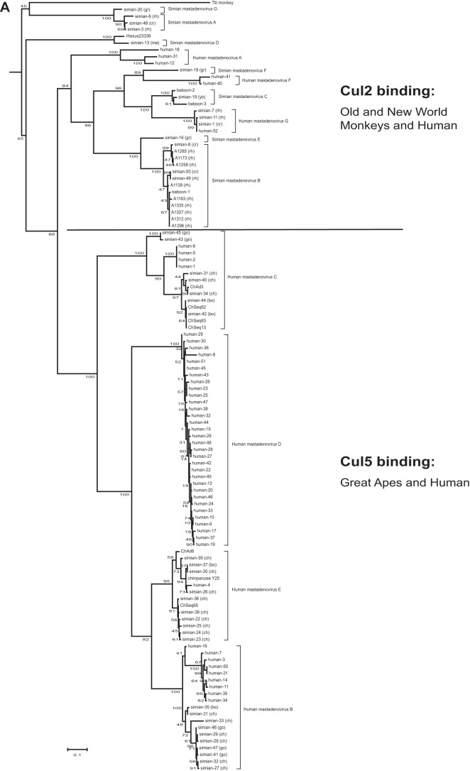
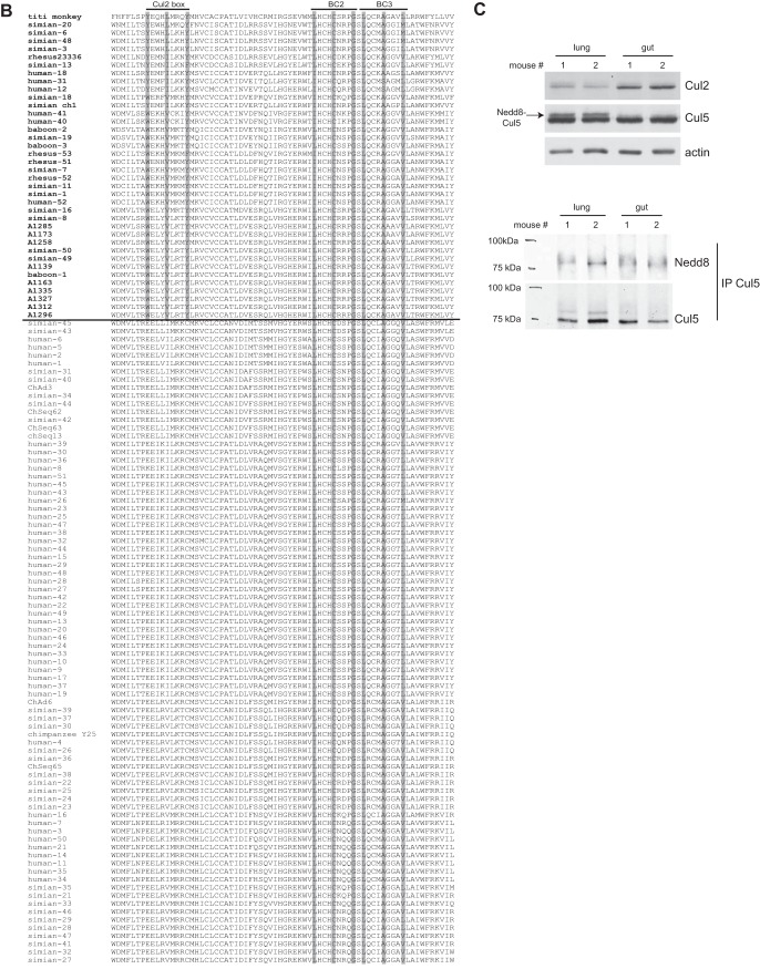
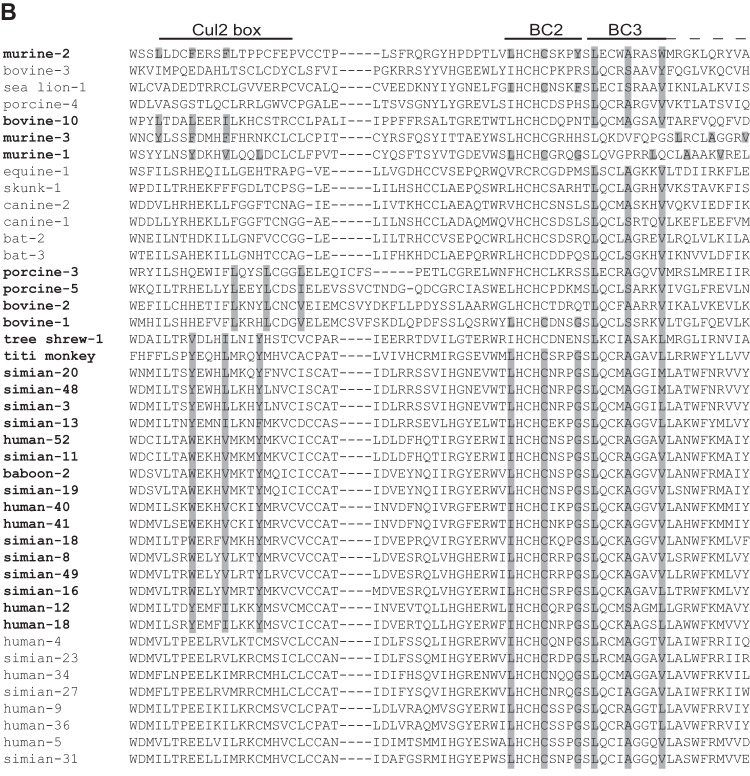
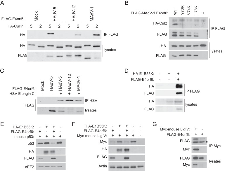
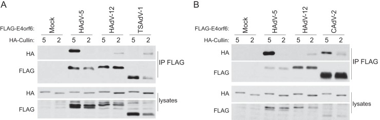
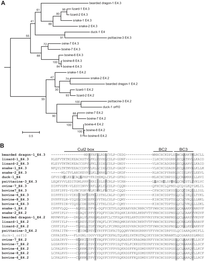
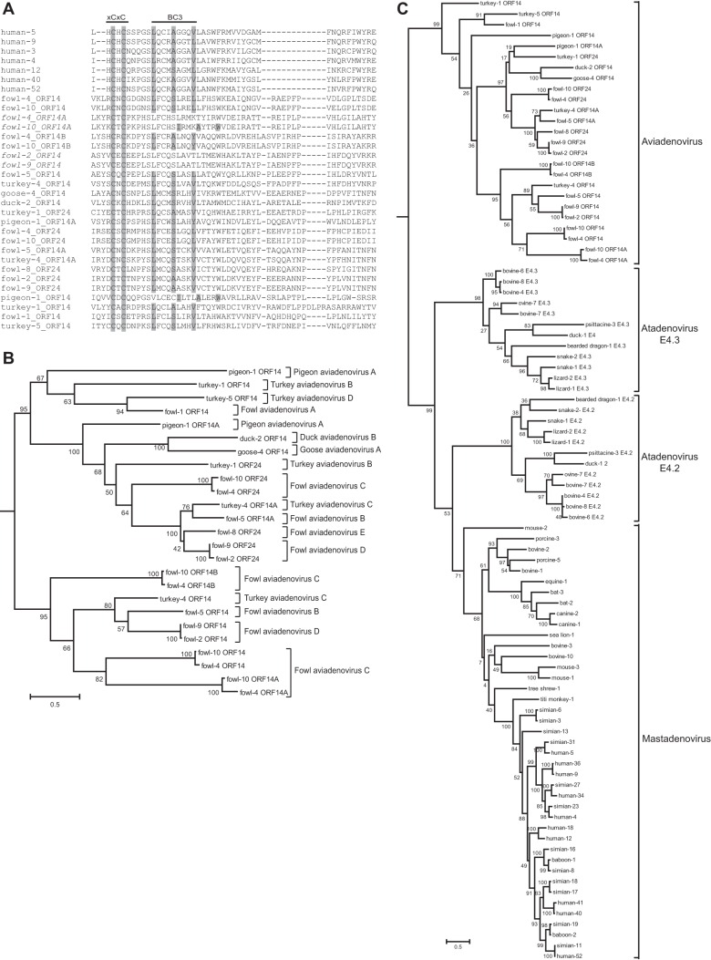
E4orf6 proteins from all human adenoviruses form Cullin-based ubiquitin ligase complexes that, in association with E1B55K, target cellular proteins for degradation. While most are assembled with Cul5, a few utilize Cul2. BC-box motifs enable all these E4orf6 proteins to assemble ligase complexes with Elongins B and C. We also identified a Cul2-box motif used for Cul2 selection in all Cul2-based complexes. With this information, we set out to determine if other adenoviruses also possess the ability to form the ligase complex and, if so, to predict their Cullin usage. Here we report that all adenoviruses known to encode an E4orf6-like protein (mastadenoviruses and atadenoviruses) maintain the potential to form the ligase complex. We could accurately predict Cullin usage for E4orf6 products of mastadenoviruses and all but one atadenovirus. Interestingly, in nonhuman primate adenoviruses, we found a clear segregation of Cullin binding, with Cul5 utilized by viruses infecting great apes and Cul2 by Old/New World monkey viruses, suggesting that a switch from Cul2 to Cul5 binding occurred during the period when great apes diverged from monkeys. Based on the analysis of Cullin selection, we also suggest that the majority of human adenoviruses, which exhibit a broader tropism for the eye and the respiratory tract, exhibit Cul5 specificity and resemble viruses infecting great apes, whereas those that infect the gastrointestinal tract may have originated from monkey viruses that share Cul2 specificity. Finally, aviadenoviruses also appear to contain E4orf6 genes that encode proteins with a conserved XCXC motif followed by, in most cases, a BC-box motif.

Importance: Two early adenoviral proteins, E4orf6 and E1B55K, form a ubiquitin ligase complex with cellular proteins to ubiquitinate specific substrates, leading to their degradation by the proteasome. In studies with representatives of each human adenovirus species, we (and others) previously discovered that some viruses use Cul2 to form the complex, while others use Cul5. In the present study, we expanded our analyses to all sequenced adenoviruses and found that E4orf6 genes from all mast- and atadenoviruses encode proteins containing the motifs necessary to form the ligase complex. We found a clear separation in Cullin specificity between adenoviruses of great apes and Old/New World monkeys, lending support for a monkey origin for human viruses of the Human mastadenovirus A, F, and G species. We also identified previously unrecognized E4orf6 genes in the aviadenoviruses that encode proteins containing motifs permitting formation of the ubiquitin ligase.


Figures

FIG 1
FIG 1
Analysis of primate adenovirus E4orf6 proteins shows a clear separation between Cul2- and Cul5-binding types. (A) Model selection by Topali for alignment of the 118 primate E4orf6 protein sequences suggested a JTT + I + G model. The PhyML calculated tree was visualized by use of Mega6 and rooted at the midpoint. Official species, if determined, are shown following the brackets. The scale bar shows an evolutionary distance of 0.1 amino acid substitution per position. The word “adenovirus” was removed from the type and strain names. Abbreviated names after the type numbers show the hosts of the simian adenoviruses, as follows: bo, bonobo; ch, chimpanzee; cr, crab-eating macaque; go, gorilla; gr, grivet; ma, macaque; rh, rhesus; yb, yellow baboon. (B) The portion of the alignment in which the Cul2 box and the BC boxes are present (roughly equivalent to the second quarter of HAdV-5 E4orf6) is shown in the same order as that for the E4orf6 proteins in the tree. The name of the virus is presented in bold if a Cul2 box is present. Residues present in Cul2 and BC boxes are shaded if the box is present. (C) (Top) Western blots of mouse lung and gut tissues were probed for Cul2, Cul5, or the actin loading control. (Bottom) The same tissue lysates were immunoprecipitated (IP) with Cul5 antibodies and the precipitates probed with Nedd8 antibodies as well as Cul5 antibodies.
FIG 1
FIG 1
Analysis of primate adenovirus E4orf6 proteins shows a clear separation between Cul2- and Cul5-binding types. (A) Model selection by Topali for alignment of the 118 primate E4orf6 protein sequences suggested a JTT + I + G model. The PhyML calculated tree was visualized by use of Mega6 and rooted at the midpoint. Official species, if determined, are shown following the brackets. The scale bar shows an evolutionary distance of 0.1 amino acid substitution per position. The word “adenovirus” was removed from the type and strain names. Abbreviated names after the type numbers show the hosts of the simian adenoviruses, as follows: bo, bonobo; ch, chimpanzee; cr, crab-eating macaque; go, gorilla; gr, grivet; ma, macaque; rh, rhesus; yb, yellow baboon. (B) The portion of the alignment in which the Cul2 box and the BC boxes are present (roughly equivalent to the second quarter of HAdV-5 E4orf6) is shown in the same order as that for the E4orf6 proteins in the tree. The name of the virus is presented in bold if a Cul2 box is present. Residues present in Cul2 and BC boxes are shaded if the box is present. (C) (Top) Western blots of mouse lung and gut tissues were probed for Cul2, Cul5, or the actin loading control. (Bottom) The same tissue lysates were immunoprecipitated (IP) with Cul5 antibodies and the precipitates probed with Nedd8 antibodies as well as Cul5 antibodies.
FIG 2
FIG 2
Analysis of E4orf6 proteins from mastadenoviruses. (A) Phylogenetic tree for the mastadenovirus E4orf6 protein sequences, including those from the 18 nonprimate viruses. For simplification of the tree, only two representatives of each primate species were included. Model selection by Topali suggested a JTT + I + G model. The PhyML calculated tree was visualized by use of Mega6 and rooted at the midpoint. Actual or predicted Cullin binding is indicted on the right, based on the presence or absence of a Cul2 box, as shown in panel B. The scale bar shows an evolutionary distance of 0.5 amino acid substitution per position. (B) The portion of the alignment in which the Cul2 box and the BC boxes are present (roughly equivalent to the second quarter of HAdV-5 E4orf6) is shown in the same order as that for the E4orf6 proteins in the tree. The name of the virus is presented in bold if a Cul2 box is present. Residues present in the Cul2 box and the BC boxes are shaded if the box is present.
FIG 2
FIG 2
Analysis of E4orf6 proteins from mastadenoviruses. (A) Phylogenetic tree for the mastadenovirus E4orf6 protein sequences, including those from the 18 nonprimate viruses. For simplification of the tree, only two representatives of each primate species were included. Model selection by Topali suggested a JTT + I + G model. The PhyML calculated tree was visualized by use of Mega6 and rooted at the midpoint. Actual or predicted Cullin binding is indicted on the right, based on the presence or absence of a Cul2 box, as shown in panel B. The scale bar shows an evolutionary distance of 0.5 amino acid substitution per position. (B) The portion of the alignment in which the Cul2 box and the BC boxes are present (roughly equivalent to the second quarter of HAdV-5 E4orf6) is shown in the same order as that for the E4orf6 proteins in the tree. The name of the virus is presented in bold if a Cul2 box is present. Residues present in the Cul2 box and the BC boxes are shaded if the box is present.
FIG 3
FIG 3
Detailed characterization of the MAdV-1 ligase complex. (A) H1299-HA-Cul2 and H1299-HA-Cul5 cells were transfected with a plasmid DNA encoding FLAG-tagged E4orf6 from the indicated virus (and HA-Cul2 for the Cul2 cell line) for 48 h. Immunoprecipitates obtained using anti-FLAG (E4orf6) antibodies and whole-cell extracts were immunoblotted as indicated, using appropriate antibodies. (B) H1299 cells were transfected with MAdV-1 FLAG-E4orf6 or a Cul2-box mutant and HA-Cul2 for 24 h. Immunoprecipitates obtained using anti-FLAG (E4orf6) antibodies and whole-cell extracts were immunoblotted as indicated, using appropriate antibodies. (C) H1299 cells were transfected with FLAG-E4orf6 from the indicated virus and HSV-tagged Elongin C for 24 h. Immunoprecipitates obtained using anti-HSV (Elongin C) antibodies and whole-cell extracts were immunoblotted as indicated, using appropriate antibodies. (D) H1299 cells were transfected with MAdV-1 FLAG-E4orf6 and HA-E1B55K, as indicated, for 24 h. Immunoprecipitates obtained using anti-HA (E1B55K) antibodies and whole-cell extracts were immunoblotted as indicated, using appropriate antibodies. (E) H1299 cells were transfected with a combination of plasmid DNAs encoding mouse p53, MAdV-1 FLAG-E4orf6, and MAdV-1 HA-E1B55K, as indicated. After 24 h, whole-cell extracts were immunoblotted as indicated, using appropriate antibodies. (F) H1299 cells were transfected and analyzed as described for panel E, except with plasmid DNA encoding mouse DNA ligase IV instead of p53. (G) H1299 cells were transfected with plasmid DNAs expressing MAdV-1 FLAG-E4orf6 and mouse DNA ligase IV, as indicated. After 24 h, immunoprecipitates obtained using anti-Myc (DNA ligase IV) antibodies and whole-cell extracts were immunoblotted as indicated, using appropriate antibodies. The asterisk denotes a background band.
FIG 4
FIG 4
E4orf6 proteins from TSAdV-1 (A) and CAdV-2 (B) bind the predicted Cullins. H1299-HA-Cul2 and H1299-HACul5 cells were transfected with a plasmid DNA encoding FLAG-tagged E4orf6 from the indicated virus (and HA-Cul2 for the Cul2 cell line) for 48 h. Immunoprecipitates obtained using anti-FLAG (E4orf6) antibodies and whole-cell extracts were immunoblotted as indicated, using appropriate antibodies.
FIG 5
FIG 5
Analysis of E4orf6 proteins from atadenoviruses. (A) Phylogenetic tree for the atadenovirus E4orf6 protein sequences. Each of the 12 atadenoviruses contained two E4orf6 genes. Model selection by Topali suggested a BLOSOM + I + G model. The PhyML calculated tree was visualized by use of Mega6 and rooted at the midpoint. The scale bar shows an evolutionary distance of 0.5 amino acid substitution per position. (B) The portion of the alignment in which the Cul2 box and the BC boxes are present (roughly equivalent to the second quarter of HAdV-5 E4orf6) is shown in the same order as that for the E4orf6 proteins in the tree. The name of the virus is presented in bold if a Cul2 box is present. Residues present in the Cul2 box and the BC boxes are shaded if the box is present.
FIG 6
FIG 6
Cullin binding prediction for atadenovirus E4orf6 proteins shows a more complex picture. H1299-HA-Cul2 and H1299-HA-Cul5 cells were transfected with a plasmid DNA encoding FLAG-tagged E4orf6 from the indicated virus (and HA-Cul2 for the Cul2 cell line). After 48 h, immunoprecipitates obtained using anti-FLAG (E4orf6) antibodies and whole-cell extracts were immunoblotted as indicated, using appropriate antibodies.
FIG 7
FIG 7
Identification of E4orf6 proteins from aviadenoviruses. (A) E4orf6 sequences were manually identified for the 13 sequenced aviadenoviruses. These sequences were aligned with seven human adenovirus sequences as a basis to compare motifs. The portion of the alignment in which the BC boxes are present (roughly equivalent to the second quarter of HAdV-5 E4orf6) is shown. Residues present in the conserved XCXC motif and the BC box are shaded if present. (B) Phylogenetic tree for E4orf6 sequences from aviadenoviruses. Model selection by Topali suggested a BLOSUM + I + G model. The PhyML calculated tree was visualized by use of Mega6 and rooted at the midpoint. Official species names are shown following brackets. The scale bar shows an evolutionary distance of 0.5 amino acid substitution per position. (C) Phylogenetic tree for E4orf6 sequences from all three genera. For simplification of the tree, only two representatives of each primate species were included. Model selection by Topali suggested a BLOSUM + I + G model. The PhyML calculated tree was visualized by use of Mega6 and rooted at the midpoint. The scale bar shows an evolutionary distance of 0.5 amino acid substitution per position.

References

    1. Benko M, Harrach B. 1998. A proposal for a new (third) genus within the family Adenoviridae. Arch Virol 143:829–837. doi: 10.1007/s007050050335. - DOI - PubMed
    1. Benko M, Harrach B. 2003. Molecular evolution of adenoviruses. Curr Top Microbiol Immunol 272:3–35. - PubMed
    1. Davison AJ, Benko M, Harrach B. 2003. Genetic content and evolution of adenoviruses. J Gen Virol 84:2895–2908. doi: 10.1099/vir.0.19497-0. - DOI - PubMed
    1. Shenk T. 1989. Oncogenesis by DNA viruses: adenovirus, p 239–257. In Weinberg RA. (ed), Oncogenes and the molecular origins of cancer, vol 18 Cold Spring Harbor Laboratory Press, Cold Spring Harbor, NY.
    1. de Jong JC, Wigand R, Kidd AH, Wadell G, Kapsenberg JG, Muzerie CJ, Wermenbol AG, Firtzlaff RG. 1983. Candidate adenoviruses 40 and 41: fastidious adenoviruses from human infant stool. J Med Virol 11:215–231. doi: 10.1002/jmv.1890110305. - DOI - PubMed

Publication types

LinkOut - more resources