

Immuno-Positron Emission Tomography with Zirconium-89-Labeled Monoclonal Antibodies in Oncology: What Can We Learn from Initial Clinical Trials?
- PMID: 27252651
- PMCID: PMC4877495
- DOI: 10.3389/fphar.2016.00131
Immuno-Positron Emission Tomography with Zirconium-89-Labeled Monoclonal Antibodies in Oncology: What Can We Learn from Initial Clinical Trials?
Abstract
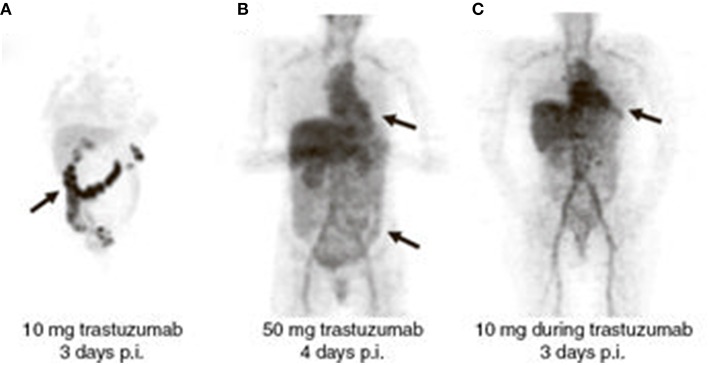
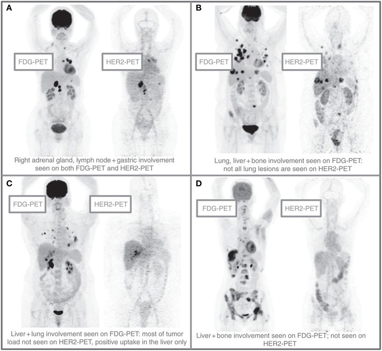
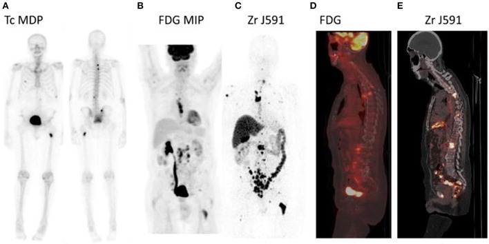
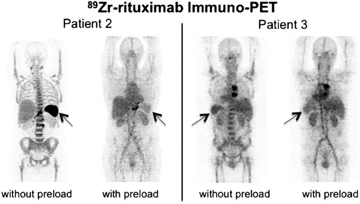
Selection of the right drug for the right patient is a promising approach to increase clinical benefit of targeted therapy with monoclonal antibodies (mAbs). Assessment of in vivo biodistribution and tumor targeting of mAbs to predict toxicity and efficacy is expected to guide individualized treatment and drug development. Molecular imaging with positron emission tomography (PET) using zirconium-89 ((89)Zr)-labeled monoclonal antibodies also known as (89)Zr-immuno-PET, visualizes and quantifies uptake of radiolabeled mAbs. This technique provides a potential imaging biomarker to assess target expression, as well as tumor targeting of mAbs. In this review we summarize results from initial clinical trials with (89)Zr-immuno-PET in oncology and discuss technical aspects of trial design. In clinical trials with (89)Zr-immuno-PET two requirements should be met for each (89)Zr-labeled mAb to realize its full potential. One requirement is that the biodistribution of the (89)Zr-labeled mAb (imaging dose) reflects the biodistribution of the drug during treatment (therapeutic dose). Another requirement is that tumor uptake of (89)Zr-mAb on PET is primarily driven by specific, antigen-mediated, tumor targeting. Initial trials have contributed toward the development of (89)Zr-immuno-PET as an imaging biomarker by showing correlation between uptake of (89)Zr-labeled mAbs on PET and target expression levels in biopsies. These results indicate that (89)Zr-immuno-PET reflects specific, antigen-mediated binding. (89)Zr-immuno-PET was shown to predict toxicity of RIT, but thus far results indicating that toxicity of mAbs or mAb-drug conjugate treatment can be predicted are lacking. So far, one study has shown that molecular imaging combined with early response assessment is able to predict response to treatment with the antibody-drug conjugate trastuzumab-emtansine, in patients with human epithelial growth factor-2 (HER2)-positive breast cancer. Future studies would benefit from a standardized criterion to define positive tumor uptake, possibly supported by quantitative analysis, and validated by linking imaging data with corresponding clinical outcome. Taken together, these results encourage further studies to develop (89)Zr-immuno-PET as a predictive imaging biomarker to guide individualized treatment, as well as for potential application in drug development.
Keywords: 89zirconium; clinical oncology; imaging biomarker; molecular imaging; monoclonal antibodies; positron emission tomography.
Figures
References
-
- Börjesson P. K., Jauw Y. W., Boellaard R., de Bree R., Comans E. F., Roos J. C., et al. . (2006). Performance of immuno-positron emission tomography with zirconium-89-labeled chimeric monoclonal antibody U36 in the detection of lymph node metastases in head and neck cancer patients. Clin. Cancer Res. 12, 2133–2140. 10.1158/1078-0432.CCR-05-2137 - DOI - PubMed
Publication types
LinkOut - more resources
Full Text Sources
Other Literature Sources
Research Materials
Miscellaneous