. 2016 May 25:7:136.
doi: 10.3389/fphar.2016.00136. eCollection 2016.

Bioabsorbable Bypass Grafts Biofunctionalised with RGD Have Enhanced Biophysical Properties and Endothelialisation Tested In vivo

Affiliations

Bioabsorbable Bypass Grafts Biofunctionalised with RGD Have Enhanced Biophysical Properties and Endothelialisation Tested In vivo

Larisa V Antonova et al. Front Pharmacol. .

Abstract

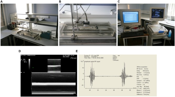
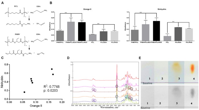
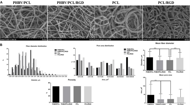
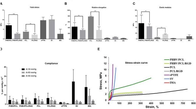
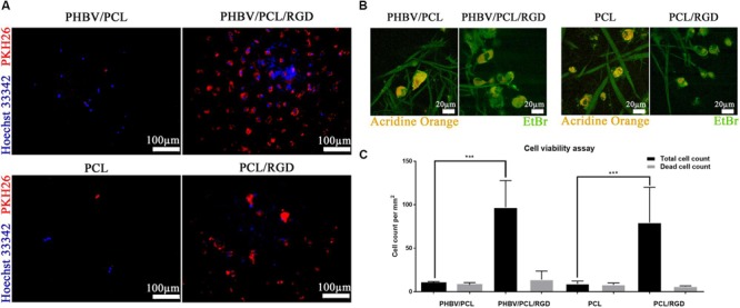
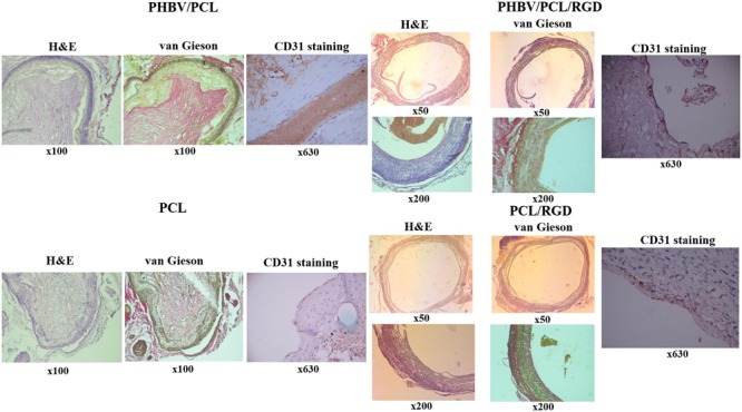
Small diameter arterial bypass grafts are considered as unmet clinical need since the current grafts have poor patency of 25% within 5 years. We have developed a 3D scaffold manufactured from natural and synthetic biodegradable polymers, poly(3-hydroxybutyrate-co-3-hydroxyvalerate) (PHBV) and poly(𝜀-caprolactone) (PCL), respectively. Further to improve the biophysical properties as well as endothelialisation, the grafts were covalently conjugated with arginine-glycine-aspartic acid (RGD) bioactive peptides. The biophysical properties as well as endothelialisation of PHBV/PCL and PCL 2 mm diameter bypass grafts were assessed with and without biofunctionalisation with RGD peptides in vitro and in vivo. Morphology of the grafts was assessed by scanning electron microscopy, whereas physico-mechanical properties were evaluated using a physiological circulating system equipped with a state of art ultrasound vascular wall tracking system. Endothelialisation of the grafts in vitro and in vivo were assessed using a cell viability assay and rat abdominal aorta replacement model, respectively. The biofunctionalisation with RGD bioactive peptides decreased mean fiber diameter and mean pore area in PHBV/PCL grafts; however, this was not the case for PCL grafts. Both PHBV/PCL and PCL grafts with RGD peptides had lower durability compared to those without; these durability values were similar to those of internal mammary artery. Modification of PHBV/PCL and PCL grafts with RGD peptides increased endothelial cell viability in vitro by a factor of eight and enhanced the formation of an endothelial cell monolayer in vivo 1 month postimplantation. In conclusion, PHBV/PCL small-caliber graft can be a suitable 3D scaffold for the development of a tissue engineering arterial bypass graft.

Keywords: RGD peptides; biocompatibility; endothelialisation; morphology; physico-mechanical properties; poly(3-hydroxybutyrate-co-3-hydroxyvalerate); poly(𝜀-caprolactone); vascular grafts.


Figures

FIGURE 1
FIGURE 1
Arterial pulsatile flow circuit. (A) Photograph of the flow circuit apparatus. (B) Detail of the perspex/saline bath and ultrasound probe in flow circuit. (C) Photograph of the ultrasound machine and wall tracking system. (D) B-mode (top) and M-mode (bottom) ultrasound images of the graft mounted in flow circuit. (E) Signal generated by anterior and posterior walls of the graft.
FIGURE 2
FIGURE 2
Modification of PHBV/PCL and PCL vascular grafts with RGD peptides. (A) Aminolysis reaction scheme between EDA, PHBV, and PCL monomers. (B) Orange II staining and ninhydrin test demonstrated increased amino group presence in PHBV/PCL and PCL grafts with EDA or RGD peptides compared to those without, data are represented as mean with standard deviation, ∗∗∗P < 0.001, two-tailed Student’s t-test. (C) Concordance test revealed a strong correlation between Orange II and ninhydrin assays for PHBV/PCL and PCL grafts with and without EDA treatment. (D) Fourier transform infrared spectroscopy showed the intensified signal from N–H stretching (around 960 cm-1, black circles) and the increase in amino residues in PHBV/PCL and PCL grafts with RGD peptides (brown and blue graphs, respectively). Peak around 1045 cm-1 (red circles) corresponds to Si-O-Si stretching, peaks around 1172 and 1240 cm-1 (green circles) correspond to C–O ester stretching, peak around 1720 cm-1 (blue circles) corresponds to C = O aliphatic ester stretching, and peaks around 2865 and 2941 cm-1 (violet circles) correspond to C–H asymmetric alkyl stretching. (E) Thin layer chromatography (TLC) spots after analyzing hydrolysed (1) PHBV/PCL, (2) PHBV/PCL/EDA, (3) PHBV/PCL/RGD, and (4) arginine and aspartic acid mixture on chromatographic silica to show the peptide content only in the RGD modified sample.
FIGURE 3
FIGURE 3
PHBV/PCL and PCL vascular grafts with RGD peptides have a distinct morphology. (A) Scanning electron microscopy images of PHBV/PCL and PCL grafts with and without RGD peptides. (B) Morphological parameters of PHBV/PCL and PCL grafts with and without RGD peptides; quantitative image analysis revealed a higher mean fiber diameter and mean pore area in PHBV/PCL compared to PCL grafts and in PHBV/PCL grafts with RGD peptides compared to those without, data are represented as mean with standard deviation, ∗∗∗P < 0.001, two-tailed Student’s t-test.
FIGURE 4
FIGURE 4
PHBV/PCL and PCL vascular grafts with RGD peptides have improved physico-mechanical properties. (A) Modification with RGD peptides decreased durability of PHBV/PCL and PCL grafts almost to the values of internal mammary artery (IMA). (B) Modification with RGD peptides reduced elasticity of PHBV/PCL and PCL grafts; however, it was still far from that of IMA. (C) Modification with RGD peptides decreased stiffness of PHBV/PCL grafts but increased stiffness of PCL grafts; for PCL grafts, stiffness was close to that of IMA. (D) Measurement of compliance found no significant differences for both PHBV/PCL and PCL grafts with and without RGD peptides. (E) Calculation of stress-strain curve of PHBV/PCL and PCL grafts with and without RGD peptides revealed it is still far from that of IMA. Data are represented as median with interquartile range, ∗∗P < 0.01, ∗∗∗P < 0.001, Mann–Whitney U-test.
FIGURE 5
FIGURE 5
PHBV/PCL and PCL scaffolds with RGD peptides improve cell viability. (A) Fluorescence microscopy showed several-fold increase in total cell count on PHBV/PCL and PCL scaffolds with RGD peptides in comparison with those without. Nuclei and cytoplasm are stained blue and red, respectively. (B) Confocal laser scanning microscopy confirmed the results obtained by fluorescence microscopy. Nuclei of the dead cells are stained orange whilst viable cells are stained green. (C) Quantitative analysis confirmed the results of microscopy analysis, data are represented as median with interquartile range, ∗∗∗P < 0.001, Mann–Whitney U-test.
FIGURE 6
FIGURE 6
Modification of PHBV/PCL and PCL vascular grafts with RGD peptides enhances formation of endothelial cell monolayer. H&E and van Gieson staining revealed a putative endothelial cell monolayer on PHBV/PCL and PCL grafts with RGD peptides but not on those without; this was further confirmed by CD31 staining, which identified a monolayer of CD31-positive cells (brown) at the inner surface of both polymer grafts with RGD peptides.

References

    1. Ahmed M., Hamilton G., Seifalian A. M. (2014). The performance of a small-calibre graft for vascular reconstructions in a senescent sheep model. Biomaterials 35 9033–9040. 10.1016/j.biomaterials.2014.07.008 - DOI - PubMed
    1. Antonova L. V., Mukhamadiyarov R. A., Mironov A. V., Burago A. Y., Velikanova E. A., Sidorova O. D., et al. (2015a). A morphological investigation of the polyhydroxybutyrate/valerate and polycaprolactone biodegradable small-diameter vascular graft biocompatibility. Genes Cells 10 71–77.
    1. Antonova L. V., Sevostyanova V. V., Seifalian A. M., Matveeva V. G., Velikanova E. A., Sergeeva E. A., et al. (2015b). Comparative in vitro testing of biodegradable vascular grafts for tissue engineering applications. Compl. Iss. Cardiovasc. Dis. 4 34–41.
    1. Causa F., Battista E., Della Moglie R., Guarnieri D., Iannone M., Netti P. A. (2010). Surface investigation on biomimetic materials to control cell adhesion: the case of RGD conjugation on PCL. Langmuir 26 9875–9884. 10.1021/la100207q - DOI - PubMed
    1. Chlupác J., Filová E., Bacáková L. (2009). Blood vessel replacement: 50 years of development and tissue engineering paradigms in vascular surgery. Physiol. Res. 58 S119–S139. - PubMed

LinkOut - more resources