Review
. 2016 May 25:4:94.
doi: 10.3389/fpubh.2016.00094. eCollection 2016.

Thinking, Walking, Talking: Integratory Motor and Cognitive Brain Function

Affiliations
Review

Thinking, Walking, Talking: Integratory Motor and Cognitive Brain Function

Gerry Leisman et al. Front Public Health. .

Abstract

In this article, we argue that motor and cognitive processes are functionally related and most likely share a similar evolutionary history. This is supported by clinical and neural data showing that some brain regions integrate both motor and cognitive functions. In addition, we also argue that cognitive processes coincide with complex motor output. Further, we also review data that support the converse notion that motor processes can contribute to cognitive function, as found by many rehabilitation and aerobic exercise training programs. Support is provided for motor and cognitive processes possessing dynamic bidirectional influences on each other.

Keywords: basal ganglia; cerebellum; cognitive processes; cognitive–motor interaction; executive function; motor processes; prefrontal cortex; premotor cortex.


Figures

Figure 1
Figure 1
Motor areas in the frontal lobe. The premotor cortex consists of the ventral premotor (PMv) and dorsal (PMd) regions. The medially located supplementary motor and presupplementary motor cortex are designated as preSMA and SMA. The regions below the superior frontal sulcus are where the premotor cortex can be found (PMv) and that part of the premotor-labeled PMd cortex located above the PMv. The boundary between the SMA and preSMA is the vertical anterior-commissural line [after Chouinard and Paus (54)].
Figure 2
Figure 2
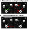
Motor network connections during unimanual hand movements in (A) healthy individuals and (B) significant differences in functional coupling during affected (right) hand movement in individuals with stroke.
Figure 3
Figure 3
Automatic and voluntary (cognitively interacting) motor control. Motor control integrates both cortical and subcortical structures principally involving those connections between the basal ganglia and frontal lobes involved in automaticity of motor function and its cognitive mediation. (8). In Parkinson’s disease, loss of dopamine in the caudal basal ganglia leads to impaired automatic movements involving circuits important in stimulus based habitual learning (red arrows) and over-reliance on cognitive components of motor control and circuits involved in reward based learning (blue arrows) (from http://neuroanatomyblog.tumblr.com).
Figure 4
Figure 4
Modes of connectivities described based either on FMRI or qEEG measurements as a basis for evaluating efficiencies of connections. The figure on the left exemplifies the fiber pathway structural connectivity, the functional connectivity (correlations), and effective connectivity (information flow) between brain regions during analysis of electrophysiological or functional imaging data. From these connectivities, we may be able to find the roles of natural phenomena as brain connectivity refers to patterns of either anatomical links, statistical dependencies, or of casual interactions among distinct areas within a nervous system. The study of the human connectome allows us to better understand the pattern of anatomical links, statistical dependencies, and/or casual interactions among distinct areas within a nervous system.
Figure 5
Figure 5
Presentation of difference maps of potential source densities (PSD) for 240–260 ms. Data for face- and leg-related words were subtracted from arm-related words. The circles on the left represent the head from above (nose = up; left = left). Circles on the right characterize lateral views on the left half of the head (nose on left). Red foci indicate stronger ingoing potentials for face-related verbs (upper diagrams) and leg-related verbs (lower diagrams). Blue foci indicate stronger ingoing activity for arm-related words. PSD is enhanced at left-lateral locations for face words and at central sites for leg words. At 240–260 ms, direct comparison of CSD topographies is provided elicited by face- and leg-related verbs. The view from the top is shown on the left and a lateral view on the left hemisphere is presented on the right. Stronger ingoing potentials for face (leg) words are specified in blue (red). Greater ingoing potentials are present at left-lateral sites for face-related items and at central sites for leg-related items [cf. Ref. (126)].
Figure 6
Figure 6
Changes in network efficiency over time post-stroke. Connectivity parameters between nodes of the motor network show increased connectivity (red lines), which are primarily seen as interhemispheric connections between M1 and contralesional sensorimotor regions. Reduced connectivity (blue lines) is mainly found in ipsilesional subcortical areas and cerebellum. IH, ipsilesional hemisphere; CH, contralesional hemisphere; M1, primary motor cortex; PCG, post-central gyrus; PMd, dorsolateral premotor cortex; PMv, ventrolateral premotor cortex; SMA, supplementary motor area; Th, thalamus; BG, basal ganglia; SPL, superior parietal lobule; SCb, superior cerebellum; DN, dentate nucleus; AICb, anterior inferior cerebellum. Reprinted with permission from Wang et al. (128).
Figure 7
Figure 7
Topographic maps of the P3 component as a function of session and task congruency on a modified Flanker task assessment of inhibitory control [cf. Ref. (145)]. Two trials were presented, one congruent and the other incongruent that necessitated participants to press a key corresponding to a centrally presented target arrow. Congruent trials contained an array of five arrows all facing in the same direction and the incongruent trials had the four contiguous arrows facing in the converse direction to the target arrow [cf. Ref. (144)].
Figure 8
Figure 8
Relationship between the total ISAT (Illinois Standards Achievement test measuring reading mathematics and science) score and the number of fitnessgram tests in which student scored in the healthy fitness zone. The Fitnessgram is a composite of aerobic capacity (PACER), muscle (strength and flexibility), and body mass index here in 259, third to fifth school children controlled for age, sex, socioeconomic status, and physical fitness [from Castelli et al. (120)].
Figure 9
Figure 9
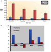
Effects of exercise on (A) cognitive performance. (B) academic skills [from Ref. (165)].
Figure 10
Figure 10
Adiposity, cognition, and academic achievement. The relationship between abdominal fat mass (in kilogram) on cognitive inhibitory control, working memory, and academic achievement (N = 122 children between the ages of 7–9 years controlled for age, sex, fitness, socioeconomic status, and IQ) [from Ref. (124)].
Figure 11
Figure 11
Walking associated with gray matter volume increases in specific brain regions. (A) Demonstrates the effects of physical activity on the brain at baseline. (B) Demonstrates the effects of walking greater than 72 streets showing greater gray matter volume.

References

    1. Harcourt-Smith WHE. The first hominins and the origins of bipedalism. Evol Educ Outreach (2010) 3(3):333–40.
    1. Falk D, Zollikofer CP, Morimoto N, Ponce de León MS. Metopic suture of Taung (Australopithecus africanus) and its implications for hominin brain evolution. Proc Nat Acad Sci U S A (2012) 109(22):8467–70.10.1073/pnas.1119752109 - DOI - PMC - PubMed
    1. Dennis MY, Nuttle X, Sudmant PH, Antonacci F, Graves TA, Nefedov M, et al. Evolution of human-specific neural SRGAP2 genes by incomplete segmental duplication. Cell (2012) 149(4):912–22.10.1016/j.cell.2012.03.033 - DOI - PMC - PubMed
    1. Melillo R, Leisman G. Neurobehavioral Disorders of Childhood: An Evolutionary Approach. New York, NY: Springer; (2009). 2009 p.
    1. Vercruyssen M, Simonton K. Effects of posture on mental performance: we think faster on our feet than on our seat. In: Lueder R, Noro K, editors. Hard Facts about Soft Machines: The Ergonomics of Seating. Bristol, PA: Taylor & Francis; (1994). p. 119–32.

LinkOut - more resources