

Clock Gene Bmal1 Modulates Human Cartilage Gene Expression by Crosstalk With Sirt1
- PMID: 27253997
- PMCID: PMC4967114
- DOI: 10.1210/en.2015-2042
Clock Gene Bmal1 Modulates Human Cartilage Gene Expression by Crosstalk With Sirt1
Abstract
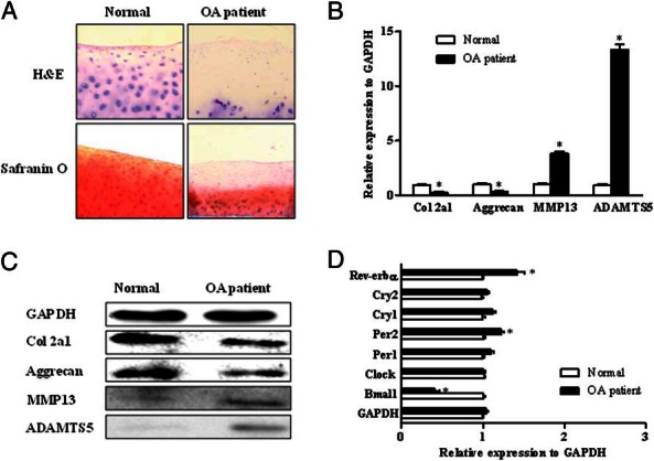
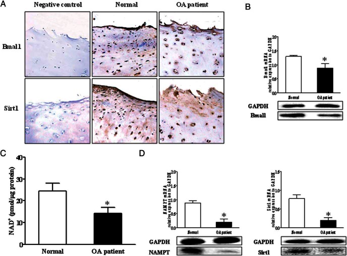
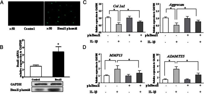
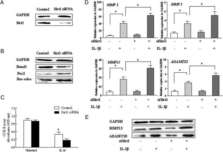
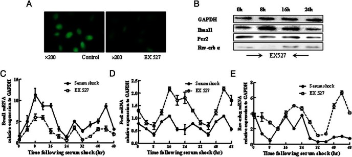
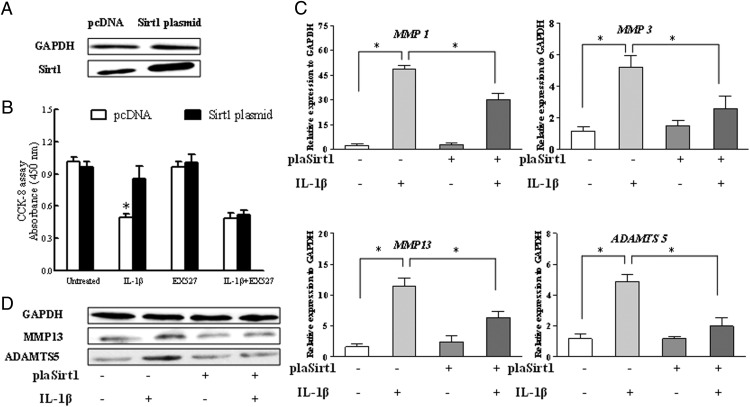
The critical regulation of the peripheral circadian gene implicated in osteoarthritis (OA) has been recently recognized; however, the causative role and clinical potential of the peripheral circadian rhythm attributable to such effects remain elusive. The purpose of this study was to elucidate the role of a circadian gene Bmal1 in human cartilage and pathophysiology of osteoarthritis. In our present study, the mRNA and protein levels of circadian rhythm genes, including nicotinamide adenine dinucleotide oxidase (NAD(+)) and sirtuin 1 (Sirt1), in human knee articular cartilage were determined. In OA cartilage, the levels of both Bmal1 and NAD(+) decreased significantly, which resulted in the inhibition of nicotinamide phosphoribosyltransferase activity and Sirt1 expression. Furthermore, the knockdown of Bmal1 was sufficient to decrease the level of NAD(+) and aggravate OA-like gene expression changes under the stimulation of IL-1β. The overexpression of Bmal1 relieved the alteration induced by IL-1β, which was consistent with the effect of the inhibition of Rev-Erbα (known as NR1D1, nuclear receptor subfamily 1, group D). On the other hand, the transfection of Sirt1 small interfering RNA not only resulted in a reduction of the protein expression of Bmal1 and a moderate increase of period 2 (per2) and Rev-Erbα but also further exacerbated the survival of cells and the expression of cartilage matrix-degrading enzymes induced by IL-1β. Overexpression of Sirt1 restored the metabolic imbalance of chondrocytes caused by IL-1β. These observations suggest that Bmal1 is a key clock gene to involve in cartilage homeostasis mediated through sirt1 and that manipulating circadian rhythm gene expression implicates an innovative strategy to develop novel therapeutic agents against cartilage diseases.
Figures

Similar articles
-
Dysregulated circadian rhythm pathway in human osteoarthritis: NR1D1 and BMAL1 suppression alters TGF-β signaling in chondrocytes.Osteoarthritis Cartilage. 2017 Jun;25(6):943-951. doi: 10.1016/j.joca.2016.11.007. Epub 2016 Nov 22. Osteoarthritis Cartilage. 2017. PMID: 27884645 Free PMC article.
-
CLOCK/BMAL1 regulates circadian change of mouse hepatic insulin sensitivity by SIRT1.Hepatology. 2014 Jun;59(6):2196-206. doi: 10.1002/hep.26992. Epub 2014 Apr 25. Hepatology. 2014. PMID: 24442997
-
IL-1β induces changes in expression of core circadian clock components PER2 and BMAL1 in primary human chondrocytes through the NMDA receptor/CREB and NF-κB signalling pathways.Cell Signal. 2021 Nov;87:110143. doi: 10.1016/j.cellsig.2021.110143. Epub 2021 Sep 3. Cell Signal. 2021. PMID: 34481895
-
Moving to the Rhythm with Clock (Circadian) Genes, Autophagy, mTOR, and SIRT1 in Degenerative Disease and Cancer.Curr Neurovasc Res. 2017;14(3):299-304. doi: 10.2174/1567202614666170718092010. Curr Neurovasc Res. 2017. PMID: 28721811 Free PMC article. Review.
-
Circadian molecular clock in lung pathophysiology.Am J Physiol Lung Cell Mol Physiol. 2015 Nov 15;309(10):L1056-75. doi: 10.1152/ajplung.00152.2015. Epub 2015 Sep 11. Am J Physiol Lung Cell Mol Physiol. 2015. PMID: 26361874 Free PMC article. Review.
Cited by
-
Fibroblast-like synoviocytes orchestrate daily rhythmic inflammation in arthritis.Open Biol. 2024 Jul;14(7):240089. doi: 10.1098/rsob.240089. Epub 2024 Jul 10. Open Biol. 2024. PMID: 38981514 Free PMC article.
-
The role of sirtuin 1 and its activator, resveratrol in osteoarthritis.Biosci Rep. 2019 May 10;39(5):BSR20190189. doi: 10.1042/BSR20190189. Print 2019 May 31. Biosci Rep. 2019. PMID: 30996115 Free PMC article. Review.
-
The therapeutic effect and mechanism of melatonin on osteoarthritis: From the perspective of non-coding RNAs.Front Genet. 2022 Oct 4;13:968919. doi: 10.3389/fgene.2022.968919. eCollection 2022. Front Genet. 2022. PMID: 36267400 Free PMC article. Review.
-
BMAL1 regulates mitochondrial homeostasis in renal ischaemia-reperfusion injury by mediating the SIRT1/PGC-1α axis.J Cell Mol Med. 2022 Apr;26(7):1994-2009. doi: 10.1111/jcmm.17223. Epub 2022 Feb 17. J Cell Mol Med. 2022. PMID: 35174626 Free PMC article.
-
Role of the Inflammation-Autophagy-Senescence Integrative Network in Osteoarthritis.Front Physiol. 2018 Jun 25;9:706. doi: 10.3389/fphys.2018.00706. eCollection 2018. Front Physiol. 2018. PMID: 29988615 Free PMC article. Review.
References
-
- Vignon E, Arlot M, Meunier P, Vignon G. Quantitative histological changes in osteoarthritic hip cartilage. Morphometric analysis of 29 osteoarthritic and 26 normal human femoral heads. Clin Orthop Relat Res. 1974(103):269–278. - PubMed
-
- Kronenberg HM. Developmental regulation of the growth plate. Nature. 2003;423(6937):332–336. - PubMed
-
- Reppert SM, Weaver DR. Weaver. Coordination of circadian timing in mammals. Nature. 2002;418(6901):935–941. - PubMed
-
- Schibler U, Sassone-Corsi P. A web of circadian pacemakers. Cell. 2002;111(7):919–922. - PubMed
-
- Fu L, Lee CC. The circadian clock: pacemaker and tumour suppressor. Nat Rev Cancer. 2003;3(5):350–361. - PubMed
Publication types
MeSH terms
Substances
Grants and funding
LinkOut - more resources
Full Text Sources
Other Literature Sources
