. 2016 Jul;12(27):3677-89.
doi: 10.1002/smll.201600178. Epub 2016 Jun 2.

Reduced Graphene Oxide-GelMA Hybrid Hydrogels as Scaffolds for Cardiac Tissue Engineering

Affiliations

Reduced Graphene Oxide-GelMA Hybrid Hydrogels as Scaffolds for Cardiac Tissue Engineering

Su Ryon Shin et al. Small. 2016 Jul.

Abstract

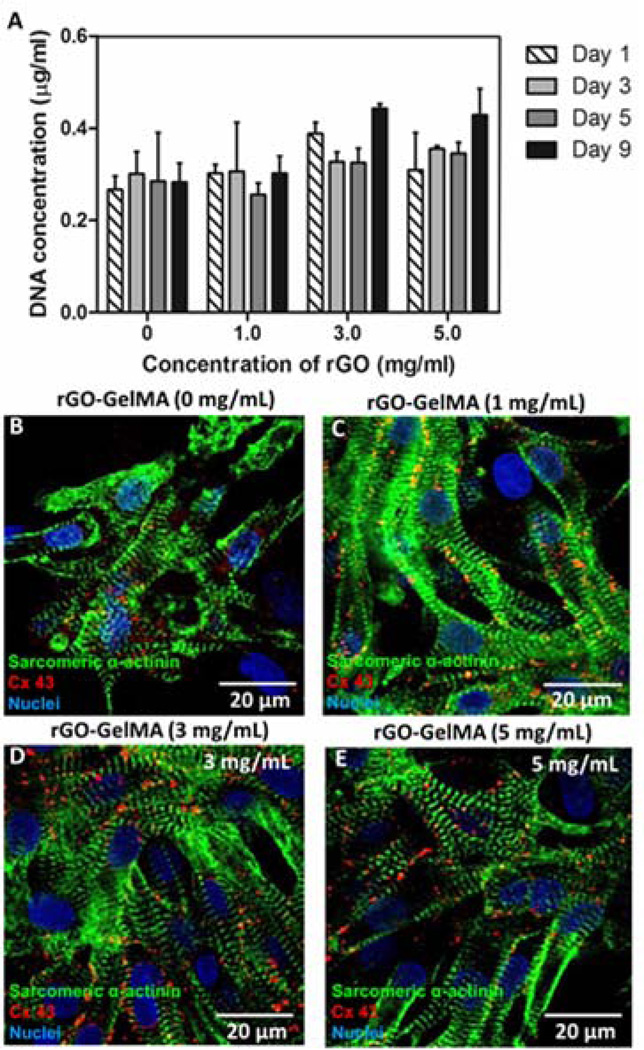
Biomaterials currently used in cardiac tissue engineering have certain limitations, such as lack of electrical conductivity and appropriate mechanical properties, which are two parameters playing a key role in regulating cardiac cell behavior. Here, the myocardial tissue constructs are engineered based on reduced graphene oxide (rGO)-incorporated gelatin methacryloyl (GelMA) hybrid hydrogels. The incorporation of rGO into the GelMA matrix significantly enhances the electrical conductivity and mechanical properties of the material. Moreover, cells cultured on composite rGO-GelMA scaffolds exhibit better biological activities such as cell viability, proliferation, and maturation compared to ones cultured on GelMA hydrogels. Cardiomyocytes show stronger contractility and faster spontaneous beating rate on rGO-GelMA hydrogel sheets compared to those on pristine GelMA hydrogels, as well as GO-GelMA hydrogel sheets with similar mechanical property and particle concentration. Our strategy of integrating rGO within a biocompatible hydrogel is expected to be broadly applicable for future biomaterial designs to improve tissue engineering outcomes. The engineered cardiac tissue constructs using rGO incorporated hybrid hydrogels can potentially provide high-fidelity tissue models for drug studies and the investigations of cardiac tissue development and/or disease processes in vitro.

Keywords: bioactuator; cardiac tissue engineering; gelatin; hydrogel; reduced graphene oxide.


Figures

Figure 1
Figure 1. Chemical characterization of the GO and rGO nanoparticles
(A) An optical image of GO and rGO dispersions. The initial brownish color of the GO solution changed to black after the reduction process. (B) FTIR spectra of GO and rGO. XPS spectra of (C & D) GO and (E & F) rGO.
Figure 2
Figure 2. Physical characterization of the GO and rGO nanoparticles
TEM images of (A) GO and (B) rGO sheets. AFM images of (C) GO and (D) GelMA coated rGO. (E & F) The height profile along the indicated line in images (C) & (D) respectively.
Figure 3
Figure 3. Structural and electrical properties of pristine GelMA and rGO-GelMA hydrogels
(A) Optical images and (B–E) SEM images of rGO-GelMA hydrogels with various concentrations of rGO: 0, 1, 3 and 5 mg• mL−1. The overall porosity of the gel were found to increase with increasing rGO concentration. (F) The elastic modulus of rGO-GelMA under compression at fully swollen state varies significantly with rGO concentration (*p < 0.05). (G) Electrical impedance curves of rGO-GelMA hydrogels with various concentrations of rGO. The impedance values were significantly lower for the samples containing rGO due to the good intrinsic conductivity of the bridging rGO sheets.
Figure 4
Figure 4. Phenotype of cardiac cells on pristine GelMA and rGO-GelMA hydrogels
(A) Cellular DNA content of cardiomyocytes on days 1, 3, 5, and 9 after seeding (n=4, avg±SD). The cellular DNA content stayed relatively constant over a period of 9 days in all samples. (B–E) Images of cardiomyocytes immunostained for cardiac markers on day 8: sarcomeric α-actinin (green), connexin 43 (red), nucleus (blue). Four different concentrations of rGO are shown: (B) 0 mg• mL−1, (C) 1 mg• mL−1, (D) 3 mg• mL−1, (E) 5 mg• mL−1. The 2D tissues on rGO-GelMA samples showed well-defined and more uniaxially aligned sarcomeric structures and more homogeneously distributed Cx-43, compared to those on pristine GelMA samples.
Figure 5
Figure 5. Electrophysiological properties of the engineered cardiac tissues
(A) Spontaneous beating rates of cardiomyocytes seeded on pristine GelMA (0 mg•mL−1) and rGO-GelMA (1, 3, 5 mg•mL−1) hydrogels as a function of incubation time. (B) Beating patterns of cardiomyocytes recorded on day 6 on rGO-GelMA hydrogels. Phase contrast images showing cultured cardiomyocytes (C) on pristine GelMA and (D) on 3 mg•mL−1 rGO-GelMA substrates 6 days after seeding. (E) Optical images show the relaxed (left) and contracted (right) cardiac tissue constructs cultured on a 3 mg•mL−1 rGO-GelMA sample stimulated using an external electrical field. (F) Response of a 3 mg•mL−1 rGO-GelMA tissue sample to an applied external electrical field at various frequencies (0.5, 1 and 2 Hz).
Figure 6
Figure 6. Function of cardiac tissues on of GO and rGO hybrid hydrogel
(A) Optical images of rGO- and GO-GelMA hydrogels with 3 mg•mL−1 rGO or GO respectively. (B) The elastic modulus of rGO- and GO-GelMA hydrogels under compression at fully swollen state. Phase contrast images showing cardiomyocytes on day 6 post seeding: (C) on GO-GelMA and (D) on rGO-GelMA. The more homogeneously distributed and aligned cardiomyocytes on the rGO-GelMA hydrogels allowed the formation of a well-organized cell sheet. (E) Cellular DNA content of cardiomyocytes on days 1 and 3 after seeding (n=4, avg±SD). The cellular DNA content stayed relatively constant over a period of 5 days in all samples. (*p < 0.05, **p < 0.001) Immunostaining of f-actin (green) and nucleus (blue) on day 2 after cell culture showed cardiomyocyte adhesion and spreading on (F) GO- and (G) rGO-GelMA hydrogel surfaces. Images of cardiomyocytes immunostained for cardiac markers with sarcomeric α-actinin (green), Cx-43 (red), and nuclei (blue) on (F) GO- and (G) rGO-GelMA hydrogels (day 8 of culture). (J) Spontaneous beating rates of cardiomyocytes seeded on GO- and rGO-GelMA hydrogels on day 5.
Schematic 1
Schematic 1. Schematic illustration of the rGO-GelMA synthesis process
(A) Process of producing rGO from GO using ascorbic acid. (B) Preparation procedure of rGO-GelMA hybrid hydrogels.

References

    1. Segers VF, Lee RT. Nature. 2008;451:937. - PubMed
    2. Alcon A, Cagavi Bozkulak E, Qyang Y. Cell Mol Life Sci. 2012;69:2635. - PMC - PubMed
    1. Coulombe KL, Bajpai VK, Andreadis ST, Murry CE. Annu Rev Biomed Eng. 2014;16:1. - PMC - PubMed
    1. Dvir T, Timko BP, Brigham MD, Naik SR, Karajanagi SS, Levy O, Jin H, Parker KK, Langer R, Kohane DS. Nat Nanotechnol. 2011;6:720. - PMC - PubMed
    1. Zhang D, Shadrin IY, Lam J, Xian HQ, Snodgrass HR, Bursac N. Biomaterials. 2013;34:5813. - PMC - PubMed
    1. Camelliti P, Borg TK, Kohl P. Cardiovasc Res. 2005;65:40. - PubMed

MeSH terms