Case Reports
. 2016 Jun 3;10(1):149.
doi: 10.1186/s13256-016-0952-1.

Septochoanal polyp with osseous metaplasia: a case report

Affiliations
Case Reports

Septochoanal polyp with osseous metaplasia: a case report

Chakapan Promsopa. J Med Case Rep. .

Abstract

Background: Polyps originating from the posterior septum with choanal extension, also known as "septochoanal polyps," are uncommon, and septochoanal polyps with central calcification are extremely rare. We report the second case of septochoanal polyps with central calcification in the English literature.

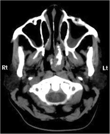
Case presentation: A 55-year-old Thai woman presented with a progressive left-side nasal obstruction. An examination of her nose revealed an irregular lobulated mass, yellow in color, with a smooth surface that arose from her posterior nasal septum and extended down to her nasopharynx. Computed tomography revealed a large choanal mass with a central ossified structure. A punch biopsy was performed and microscopic examination showed an inflammatory polyp. The mass was removed using an endoscopic surgery technique, and the histology of this lesion confirmed a typical presentation of choanal polyps.

Conclusions: Although septochoanal polyps with osseous metaplasia are known to be very rare, physicians should be aware of them and include them in the differential diagnosis of choanal mass with central calcification lesions.

Keywords: Calcification; Choanal; Osseous metaplasia; Polyp; Septochoanal.


Figures

Fig. 1
Fig. 1
Axial view computed tomography soft tissue window demonstrated a 5×3×3 cm lobulated non-enhancing mass in posterior nasal cavity and nasopharynx arising from the left posterior nasal septum. Dense central calcification is noted. Lt left, Rt right
Fig. 2
Fig. 2
Surgical specimen. a The choanal polyp measured 5×3×3 cm. The irregular lobulated mass was white-yellow in color. The pedicle is shown at the upper end (arrow). b The mass contains central osseous metaplasia (*)
Fig. 3
Fig. 3
Histopathology (hematoxylin and eosin; original magnification, ×40) revealed mature trabecular bone tissue (arrows) covered with respiratory polypoid mucosa without eosinophils

References

    1. Lopatin A, Bykova V, Piskunov G. Choanal polyps: one entity, one surgical approach. Rhinology. 1997;35:79–83. - PubMed
    1. Ozgirgin ON, Kutluay L, Akkuzu G, et al. Choanal polyp originating from the nasal septum: a case report. Am J Otolaryngol. 2003;24:261–4. doi: 10.1016/S0196-0709(03)00048-6. - DOI - PubMed
    1. Chaiyasate S, Roongrotwattanasiri K, Patumanond J, et al. Antrochoanal polyps: how long should follow-up be after surgery. Int J Otolaryngol. 2015;2015:1–5. - PMC - PubMed
    1. Choudhury N, Hariri A, Saleh H. Endoscopic management of antrochoanal polyps: a single UK centre’s experience. Eur Arch Otorhinolaryngol. 2015;272(9):2305–11. doi: 10.1007/s00405-014-3163-7. - DOI - PubMed
    1. Birkent H, Karahatay S, Durmaz A, et al. Choanal polyp originating from the nasal septum: septochoanal polyp. Kulak Burun Bogaz Ihtis Derg. 2009;19(3):163–6. - PubMed

Publication types

LinkOut - more resources