. 2016 Jul:43:79-88.
doi: 10.1016/j.neurobiolaging.2016.03.026. Epub 2016 Apr 6.

Age-dependent differences in brain tissue microstructure assessed with neurite orientation dispersion and density imaging

Affiliations

Age-dependent differences in brain tissue microstructure assessed with neurite orientation dispersion and density imaging

Andrew P Merluzzi et al. Neurobiol Aging. 2016 Jul.

Abstract

Human aging is accompanied by progressive changes in executive function and memory, but the biological mechanisms underlying these phenomena are not fully understood. Using neurite orientation dispersion and density imaging, we sought to examine the relationship between age, cellular microstructure, and neuropsychological scores in 116 late middle-aged, cognitively asymptomatic participants. Results revealed widespread increases in the volume fraction of isotropic diffusion and localized decreases in neurite density in frontal white matter regions with increasing age. In addition, several of these microstructural alterations were associated with poorer performance on tests of memory and executive function. These results suggest that neurite orientation dispersion and density imaging is capable of measuring age-related brain changes and the neural correlates of poorer performance on tests of cognitive functioning, largely in accordance with published histological findings and brain-imaging studies of people of this age range. Ultimately, this study sheds light on the processes underlying normal brain development in adulthood, knowledge that is critical for differentiating healthy aging from changes associated with dementia.

Keywords: Aging; Cognition; Diffusion-weighted imaging; MRI; Microstructure; Neurites.


Figures

Figure 1
Figure 1
Shown here are representative brain maps from one participant. Neurite density (panel A), orientation dispersion (panel B), isotropic volume fraction (panel C), fractional anisotropy (panel D), and mean diffusivity (panel E) are displayed.
Figure 2
Figure 2
Shown here is the relationship between age (in years) and neurite density (NDI) in four white matter regions in the frontal cortex, including bilateral ventromedial and dorsomedial frontal cortex. In all cases, age is negatively associated with neurite density, accounting for sex, APOE ε4 genotype, and parental history of AD.
Figure 3
Figure 3
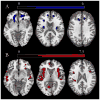
Axial view brain maps with highlighted regions in which neurite density (NDI) decreases with increasing age in WM (panel A) and regions in which the volume fraction of isotropic diffusion (Viso) increases with increasing age in GM (panel B). All images are presented in neurological convention (left on the left), and results are family-wise error corrected with a threshold of p < 0.05. The color bars represent the size of the t-statistic, with blue representing decreased NDI and red representing increased Viso.
Figure 4
Figure 4
Panel A: Lower neurite density (NDI) in left frontal superior white matter (WM) is associated with poorer performance on the learning portion of the Rey Auditory Verbal Learning Test (RAVLT). Panel B: Lower NDI in right frontal superior WM is associated with poorer performance on the trail-making test (TMT) part B (this scatterplot excludes two participants who scored poorly on this test; n=114). Panel C: Lower NDI in left frontal inferior WM is associated with poorer performance on the learning portion of the RAVLT. Panel D: Lower NDI in right frontal inferior WM is associated with poorer performance on the learning portion of the RAVLT. Corresponding statistical brain maps in the transverse plane are presented to the right of the scatterplots, with highlighted regions in which NDI is reduced with age in bilateral dorsomedial (top) and ventromedial (bottom) frontal cortex. All neuropsychological results account for age, sex, APOE ε4 genotype, years of education, and parental history of AD.

References

    1. Aboitiz F, Rodriquez E, Olivares R, Zaidel E. Age-related changes in the fibre composition of the human corpus callosum: sex differences. NeuroReport. 1996;7(11):1761–4. - PubMed
    1. Adluru G, Gur Y, Anderson JS, Richards LG, Adluru N, DiBella EV. Assessment of white matter microstructure in stroke patients using NODDI. Engineering in Medicine and Biology Society (EMBC), 2014 36th Annual International Conference of the IEEE; IEEE; 2014. pp. 742–5. - PMC - PubMed
    1. Alexander MP, Stuss DT, Picton T, Shallice T, Gillingham S. Regional frontal injuries cause distinct impairments in cognitive control. Neurology. 2007;68(18):1515–23. - PubMed
    1. Allen JS, Bruss J, Brown CK, Damasio H. Normal neuroanatomical variation due to age: the major lobes and a parcellation of the temporal region. Neurobiol Aging. 2005;26(9):1245–60. doi: 10.1016/j.neurobiolaging.2005.05.023. discussion 79–82. - DOI - PubMed
    1. Anderson B, Rutledge V. Age and hemisphere effects on dendritic structure. Brain. 1996;119:1983–90. - PubMed

Publication types