

Interactions between amiodarone and the hERG potassium channel pore determined with mutagenesis and in silico docking
- PMID: 27256139
- PMCID: PMC4959829
- DOI: 10.1016/j.bcp.2016.05.013
Interactions between amiodarone and the hERG potassium channel pore determined with mutagenesis and in silico docking
Abstract
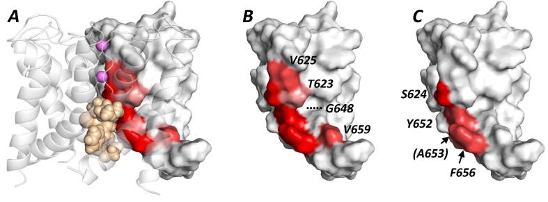
The antiarrhythmic drug amiodarone delays cardiac repolarisation through inhibition of hERG-encoded potassium channels responsible for the rapid delayed rectifier potassium current (IKr). This study aimed to elucidate molecular determinants of amiodarone binding to the hERG channel. Whole-cell patch-clamp recordings were made at 37°C of ionic current (IhERG) carried by wild-type (WT) or mutant hERG channels expressed in HEK293 cells. Alanine mutagenesis and ligand docking were used to investigate the roles of pore cavity amino-acid residues in amiodarone binding. Amiodarone inhibited WT outward IhERG tails with a half-maximal inhibitory concentration (IC50) of ∼45nM, whilst inward IhERG tails in a high K(+) external solution ([K(+)]e) of 94mM were blocked with an IC50 of 117.8nM. Amiodarone's inhibitory action was contingent upon channel gating. Alanine-mutagenesis identified multiple residues directly or indirectly involved in amiodarone binding. The IC50 for the S6 aromatic Y652A mutation was increased to ∼20-fold that of WT IhERG, similar to the pore helical mutant S624A (∼22-fold WT control). The IC50 for F656A mutant IhERG was ∼17-fold its corresponding WT control. Computational docking using a MthK-based hERG model differentiated residues likely to interact directly with drug and those whose Ala mutation may affect drug block allosterically. The requirements for amiodarone block of aromatic residues F656 and Y652 within the hERG pore cavity are smaller than for other high affinity IhERG inhibitors, with relative importance to amiodarone binding of the residues investigated being S624A∼Y652A>F656A>V659A>G648A>T623A.
Keywords: Amiodarone; Amiodarone hydrochloride (PubChem CID: 441325); Antiarrhythmic; I(Kr); Long QT; QT interval; hERG.
Copyright © 2016 The Author(s). Published by Elsevier Inc. All rights reserved.
Figures


Similar articles
-
Molecular determinants of hERG potassium channel inhibition by disopyramide.J Mol Cell Cardiol. 2012 Jan;52(1):185-95. doi: 10.1016/j.yjmcc.2011.09.021. Epub 2011 Sep 29. J Mol Cell Cardiol. 2012. PMID: 21989164
-
Ranolazine inhibition of hERG potassium channels: drug-pore interactions and reduced potency against inactivation mutants.J Mol Cell Cardiol. 2014 Sep;74(100):220-30. doi: 10.1016/j.yjmcc.2014.05.013. Epub 2014 May 27. J Mol Cell Cardiol. 2014. PMID: 24877995 Free PMC article.
-
Molecular basis of hERG potassium channel blockade by the class Ic antiarrhythmic flecainide.J Mol Cell Cardiol. 2015 Sep;86:42-53. doi: 10.1016/j.yjmcc.2015.06.021. Epub 2015 Jul 6. J Mol Cell Cardiol. 2015. PMID: 26159617 Free PMC article.
-
Relationship among amiodarone, new class III antiarrhythmics, miscellaneous agents and acquired long QT syndrome.Cardiol J. 2008;15(3):209-19. Cardiol J. 2008. PMID: 18651412 Review.
-
Structural determinants for high-affinity block of hERG potassium channels.Novartis Found Symp. 2005;266:136-50; discussion 150-8. Novartis Found Symp. 2005. PMID: 16050266 Review.
Cited by
-
Cardamonin, a Novel Antagonist of hTRPA1 Cation Channel, Reveals Therapeutic Mechanism of Pathological Pain.Molecules. 2016 Aug 29;21(9):1145. doi: 10.3390/molecules21091145. Molecules. 2016. PMID: 27589700 Free PMC article.
-
Molecular determinants of HERG potassium channel blockade by domiphen bromide and benzethonium chloride.Pflugers Arch. 2025 Aug;477(8):1119-1130. doi: 10.1007/s00424-025-03104-5. Epub 2025 Jul 21. Pflugers Arch. 2025. PMID: 40685478
-
Dronedarone blockage of the tumor-related Kv10.1 channel: a comparison with amiodarone.Pflugers Arch. 2020 Jan;472(1):75-87. doi: 10.1007/s00424-019-02342-8. Epub 2020 Jan 2. Pflugers Arch. 2020. PMID: 31897736
-
Computational Modeling of Electrophysiology and Pharmacotherapy of Atrial Fibrillation: Recent Advances and Future Challenges.Front Physiol. 2018 Sep 4;9:1221. doi: 10.3389/fphys.2018.01221. eCollection 2018. Front Physiol. 2018. PMID: 30233399 Free PMC article. Review.
-
Calculation of absolute binding free energies between the hERG channel and structurally diverse drugs.Sci Rep. 2019 Nov 12;9(1):16586. doi: 10.1038/s41598-019-53120-6. Sci Rep. 2019. PMID: 31719645 Free PMC article.
References
-
- Kodama I., Kamiya K., Toyama J. Cellular electropharmacology of amiodarone. Cardiovasc. Res. 1997;35:13–29. - PubMed
-
- Doggrell S.A. Amiodarone – waxed and waned and waxed again. Expert Opin. Pharmacother. 2001;2:1877–1890. - PubMed
-
- Camm A.J., Lip G.Y., De Caterina R., Savelieva I., Atar D., Hohnloser S.H. 2012 focused update of the ESC guidelines for the management of atrial fibrillation: an update of the 2010 ESC guidelines for the management of atrial fibrillation – developed with the special contribution of the European Heart Rhythm Association. Europace. 2012;14(2012):1385–1413. - PubMed
-
- Marinelli A., Capucci A. Amiodarone (Nexterone) injection for the treatment and prophylaxis of frequently recurring ventricular fibrillation. Expert Opin. Pharmacother. 2012;13:573–584. - PubMed
Publication types
MeSH terms
Substances
Grants and funding
LinkOut - more resources
Full Text Sources
Other Literature Sources
