. 2016 Dec;2(1):54.
doi: 10.1186/s40792-016-0181-2. Epub 2016 Jun 3.

Flap revascularization in patients following immediate reconstruction using an autologous free dermal fat graft for breast cancer: a report of two cases

Affiliations

Flap revascularization in patients following immediate reconstruction using an autologous free dermal fat graft for breast cancer: a report of two cases

Hiroaki Shima et al. Surg Case Rep. 2016 Dec.

Abstract

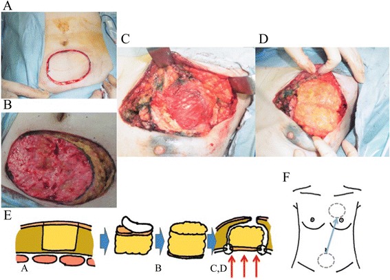
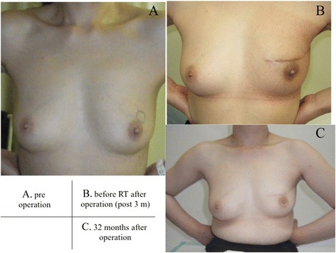
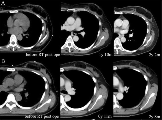
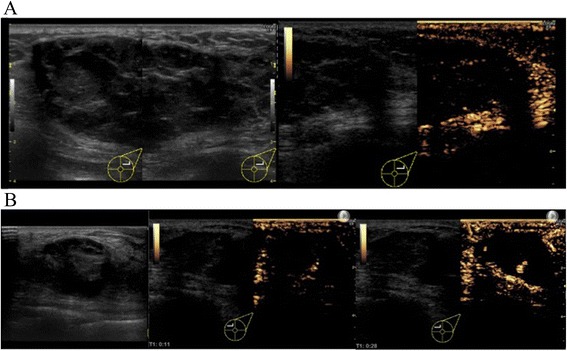
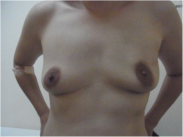
It has been reported that use of the free dermal fat graft (FDFG) technique produces a good cosmetic outcome for breast cancer. An FDFG is harvested from the lower abdomen as a columnar-shaped specimen and implanted into the defect of the breast after a partial mastectomy as a volume replacement technique. In this report, two patients who underwent breast-conserving surgery with immediate reconstruction using an autologous FDFG are described in order to show the difference in status between one case with and one without blood flow in the graft. To assess the benefit of this technique using FDFGs, their cosmetic satisfaction was evaluated using a questionnaire, graft shrinkage was measured by CT, and blood flow was assessed using contrast-enhanced ultrasound (CEUS). Both patients scored 10 of 12 points on the questionnaire. After 2 years, shrinkage of the grafts was 21.6 and 25.2 %, respectively. Although one patient had no blood flow in the center of the graft, the other had blood flow from the pectoralis major muscle to the center of the graft. While satisfaction and graft shrinkage were similar in the two patients, one case showed blood flow and had a somewhat softer graft than the other. The graft status was maintained with a good cosmetic outcome for 3 years after breast-conserving surgery with immediate reconstruction using an autologous FDFG, despite mild shrinkage and hardness of the graft. It is notable that blood flow was observed into the graft on CEUS, and more distinct perfusion was seen in the softer graft case after more than 3 years.

Keywords: Breast reconstruction; Breast-conserving surgery; Contrast-enhanced ultrasound; Tissue transplant.


Figures

Fig. 1
Fig. 1
Ultrasound (US: left panel), magnetic resonance imaging (MRI: center panel), and pathological findings (right panel) of case 1 (a, upper line) and case 2 (b, lower line). US and MRI show a localized mass in each case. Partial mastectomy was performed prior to immediate reconstruction using a free dermal fat graft, which resulted in surgical margins that were negative for cancer in both cases (a, b, right panel)
Fig. 2
Fig. 2
A free dermal fat graft (FDFG: 7.0 cm × 9.0 cm) was harvested from the lower abdomen (a). The specimen is denuded of epidermal skin and made into a columnar shape (b). The breast defect (c) was repaired and filled with the FDFG, with the denuded surface fitted to the pectoralis major muscle (d). In the semi-sitting position, nipple and breast symmetry are checked carefully. A schematic diagram of this process is shown (e, f)
Fig. 3
Fig. 3
Pictures indicated temporal statuses of Case 1; a status before operation (a), post-operative status before radiation therapy (3 months after operation) (b), and 32 months after operation (c)
Fig. 4
Fig. 4
Chronological changes of grafts in case 1 (a, upper line) and case 2 (b, lower line). After 2 years and more, shrinkage of grafts was 21.6 and 25.2 %, respectively. Slight enhancement in the center of or around the graft is indistinct in each case, and it is difficult to assess the blood flow in detail
Fig. 5
Fig. 5
Ultrasound (US) and contrast-enhanced ultrasound (CEUS) were performed in case 1 (a, upper line) and case 2 (b, lower line) over 3 years after surgery. In case 1, the graft is detected as a circumscribed oval and heterogeneous low echoic mass on US. This mass is coated by a smooth capsule with a high echogenic structure (a, left panel). CEUS shows no blood flow into the mass from the outside fat tissue (a, right panel). In case 2, the graft is seen as a circumscribed oval and heterogeneous low echoic mass on US, very similar to case 1. CEUS shows blood flow into the graft mainly from the retro-mammary fat side, which begins approximately 10 s after intravenous administration of contrast agent (center panel). In the mass, several blood flows are observed with persistent enhancement without attenuation once the vessels are enhanced for 30 s after administration (right panel)
Fig. 6
Fig. 6
Postoperative appearance of case 2. A 37-year-old woman with a localized 1.2-cm tumor in the upper region of the left breast 44 months after surgery

References

    1. Fisher B, Anderson S, Bryant J, Margolese RG, Deutsch M, Fisher ER, et al. Twenty-year follow-up of a randomized trial comparing total mastectomy, lumpectomy, and lumpectomy plus irradiation for the treatment of invasive breast cancer. N Engl J Med. 2002;347(16):1233–1241. doi: 10.1056/NEJMoa022152. - DOI - PubMed
    1. Veronesi U, Cascinelli N, Mariani L, Greco M, Saccozzi R, Luini A, et al. Twenty-year follow-up of a randomized study comparing breast-conserving surgery with radical mastectomy for early breast cancer. N Engl J Med. 2002;347(16):1227–1232. doi: 10.1056/NEJMoa020989. - DOI - PubMed
    1. Urban C, Lima R, Schunemann E, Spautz C, Rabinovich I, Anselmi K. Oncoplastic principles in breast conserving surgery. Breast. 2011;20(Suppl 3):S92–S95. doi: 10.1016/S0960-9776(11)70302-2. - DOI - PubMed
    1. Kijima Y, Yoshinaka H, Funasako Y, Kaneko K, Hirata M, Mizoguchi T, et al. Immediate breast reconstruction using autologous free dermal fat grafts provides better cosmetic results for patients with upper inner cancerous lesions. Surg Today. 2011;41(4):477–489. doi: 10.1007/s00595-010-4307-z. - DOI - PubMed
    1. Kijima Y, Yoshinaka H, Owaki T, Funasako Y, Aikou T. Immediate reconstruction using inframammary adipofascial flap of the anterior rectus sheath after partial mastectomy. Am J Surg. 2007;193(6):789–791. doi: 10.1016/j.amjsurg.2006.06.052. - DOI - PubMed

LinkOut - more resources