A novel set-up for the ex vivo analysis of mechanical properties of mouse aortic segments stretched at physiological pressure and frequency
- PMID: 27256450
- PMCID: PMC5088227
- DOI: 10.1113/JP272623
A novel set-up for the ex vivo analysis of mechanical properties of mouse aortic segments stretched at physiological pressure and frequency
Abstract
Key points: Cyclic stretch is known to alter intracellular pathways involved in vessel tone regulation. We developed a novel set-up that allows straightforward characterization of the biomechanical properties of the mouse aorta while stretched at a physiological heart rate (600 beats min-1 ). Active vessel tone was shown to have surprisingly large effects on isobaric stiffness. The effect of structural vessel wall alterations was confirmed using a genetic mouse model. This set-up will contribute to a better understanding of how active vessel wall components and mechanical stimuli such as stretch frequency and amplitude regulate aortic mechanics.
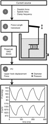
Abstract: Cyclic stretch is a major contributor to vascular function. However, isolated mouse aortas are frequently studied at low stretch frequency or even in isometric conditions. Pacing experiments in rodents and humans show that arterial compliance is stretch frequency dependent. The Rodent Oscillatory Tension Set-up to study Arterial Compliance is an in-house developed organ bath set-up that clamps aortic segments to imposed preloads at physiological rates up to 600 beats min-1 . The technique enables us to derive pressure-diameter loops and assess biomechanical properties of the segment. To validate the applicability of this set-up we aimed to confirm the effects of distension pressure and vascular smooth muscle tone on arterial stiffness. At physiological stretch frequency (10 Hz), the Peterson modulus (EP ; 293 (10) mmHg) for wild-type mouse aorta increased 22% upon a rise in pressure from 80-120 mmHg to 100-140 mmHg, while, at normal pressure, EP increased 80% upon maximal contraction of the vascular smooth muscle cells. We further validated the method using a mouse model with a mutation in the fibrillin-1 gene and an endothelial nitric oxide synthase knock-out model. Both models are known to have increased arterial stiffness, and this was confirmed using the set-up. To our knowledge, this is the first set-up that facilitates the study of biomechanical properties of mouse aortic segments at physiological stretch frequency and pressure. We believe that this set-up can contribute to a better understanding of how cyclic stretch frequency, amplitude and active vessel wall components influence arterial stiffening.
Keywords: VSMC tone; arterial stiffness; basal nitric oxide; cyclic stretch.
© 2016 The Authors. The Journal of Physiology © 2016 The Physiological Society.
Figures


References
-
- Barra JG, Armentano RL, Levenson J, Fischer EI, Pichel RH & Simon A (1993). Assessment of smooth muscle contribution to descending thoracic aortic elastic mechanics in conscious dogs. Circ Res 73, 1040–1050. - PubMed
-
- Bellien J, Favre J, Iacob M, Gao J, Thuillez C, Richard V & Joannidès R (2010). Arterial stiffness is regulated by nitric oxide and endothelium‐derived hyperpolarizing factor during changes in blood flow in humans. Hypertension 55, 674–680. - PubMed
-
- Benetos A, Safar M, Rudnichi A, Smulyan H, Richard JL, Ducimetieère P & Guize L (1997). Pulse pressure: a predictor of long‐term cardiovascular mortality in a French male population. Hypertension 30, 1410–1415. - PubMed
-
- Bia D, Aguirre I, Zócalo Y, Devera L, Cabrera Fischer E & Armentano R (2005). Regional differences in viscosity, elasticity, and wall buffering function in systemic arteries: pulse wave analysis of the arterial pressure‐diameter relationship. Rev Esp Cardiol 58, 167–174. - PubMed
-
- Boutouyrie P, Bézie Y, Lacolley P, Challande P, Chamiot‐Clerc P, Benetos A, de la Faverie JF, Safar M & Laurent S (1997). In vivo/in vitro comparison of rat abdominal aorta wall viscosity. Influence of endothelial function. Arterioscler Thromb Vasc Biol 17, 1346–1355. - PubMed
Publication types
MeSH terms
LinkOut - more resources
Full Text Sources
Other Literature Sources
Miscellaneous

