. 2016 Sep;30(9):3181-93.
doi: 10.1096/fj.201600375R. Epub 2016 Jun 2.

Resolvin D2 decreases TLR4 expression to mediate resolution in human monocytes

Affiliations

Resolvin D2 decreases TLR4 expression to mediate resolution in human monocytes

Amanda Croasdell et al. FASEB J. 2016 Sep.

Abstract

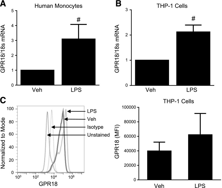
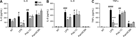
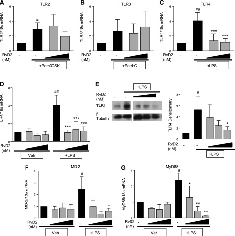
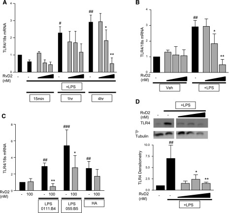
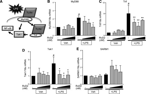
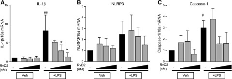
TLRs are critical for innate immunity, but excessive activation can lead to tissue damage and disease. Specialized proresolving mediators (SPMs), including resolvin D2 (RvD2), promote the active resolution of inflammation. How SPMs regulate early LPS signaling, including activation of TLR4, is unknown. We treated human THP-1 monocytic cells and primary human blood monocytes with RvD2 and LPS to evaluate modulation of TLRs. miRNA-146a overexpression and inhibition were used to dissect the mechanism of RvD2-mediated actions. We validated our studies using ELISAs for cytokines, PCR, Western blot analysis, and flow cytometry. Cells treated with 0.1% ethanol (control for RvD2) and/or PBS (control for LPS), and control microRNA mimics and inhibitors were used as controls. RvD2 reduced LPS-induced cytokines and TLR4 expression in human monocytes by up to 75%. In THP-1 cells, RvD2 reduced expression of TLR4, lymphocyte antigen 96 (MD-2), and downstream signals (MyD88, TRIF, and TAK1). These effects were partially mediated through RvD2 induction of microRNA-146a, and RvD2's actions were blocked by microRNA-146a inhibition. These new findings reveal the ability of RvD2 to reduce TLR4 expression and attenuate LPS-induced inflammation, providing a new area of SPM activity to investigate in this major area of therapeutic research.-Croasdell, A., Sime, P. J., Phipps, R. P. Resolvin D2 decreases TLR4 expression to mediate resolution in human monocytes.

Keywords: LPS; MD-2; SPM; inflammation; microRNA-146a.


Figures

Figure 1.
Figure 1.
RvD2 receptor GPR18 is increased with LPS treatment. Human blood monocytes (A) and THP-1 cells (B, C) were treated with 20 ng/ml LPS. At 4 h after activation, levels of GPR18 mRNA were assessed by RT-PCR (A, B), or protein expression was determined by flow cytometry (C). Statistical significance was determined by Student's t test, n = 3–4 independent experiments or individual donors. #P < 0.05 compared to vehicle.
Figure 2.
Figure 2.
RvD2 dampens LPS-induced cytokine production by human monocytes. Human blood monocytes were treated with 10 or 100 nM RvD2 followed by 20 ng/ml LPS, poly(I:C), or Pam3CSK. At 24 h, activation levels of IL-6 (A), IL-8 (B), and TNF-α (C) were assessed by ELISA. Statistical significance was determined by 2-way ANOVA, n = 3–4 individual donors. #P < 0.05, ##P < 0.01, ###P < 0.001, ####P < 0.0001 compared with vehicle/vehicle; *P < 0.05, **P < 0.01, ***P < 0.001 compared with activator/vehicle.
Figure 3.
Figure 3.
RvD2 dampens LPS-induced TLR4 expression in human monocytes. Human blood monocytes were treated with 10 or 100 nM RvD2 followed by 20 ng/ml LPS, poly(I:C), or Pam3CSK. AC) TLR2 (A), TLR3 (B), and TLR4 (C) mRNA expression were determined 1 h after activator stimulation by RT-PCR. DG) Effects of 1–100 nM RvD2 alone and with LPS treatment on TLR4 (D), MD-2 (F), and MyD88 (G) mRNA were also assessed; TLR4 protein expression was determined by Western blot analysis 1 h after LPS exposure (E) (representative image shown, densitometry for n = 4). Statistical significance was determined by 2-way ANOVA, n = 4–5 individual donors. #P < 0.05, ##P < 0.01 compared with vehicle/vehicle; *P < 0.05, **P < 0.01, ***P < 0.001 compared with activator/vehicle.
Figure 4.
Figure 4.
RvD2 decreases TLR4 expression in time-dependent manner in THP-1 monocytic cells. A) THP-1 cells were treated with 1–100 nM RvD2 followed by 20 ng/ml LPS. TLR4 mRNA was assessed 15 min, 1 h, and 4 h after LPS stimulation. B) Effects of 1–100 nM RvD2 alone and with LPS treatment on TLR4 mRNA were also assessed 4 h after LPS exposure (same data shown as that for vehicle/vehicle, vehicle/LPS, and RvD2/LPS groups). C) THP-1 cells were also treated with 100 nM RvD2 followed by 20 ng/ml LPS (either 0111:B4 or 055:B5) or hyaluronan, and TLR4 mRNA was assessed after 4 h. D) TLR4 protein expression was determined by Western blot analysis 4 h after LPS exposure (representative image shown, densitometry for n = 4). Statistical significance was determined by 2-way ANOVA, n = 3–5 independent experiments. #P < 0.05, ##P < 0.01, ###P < 0.001 compared with vehicle/vehicle; *P < 0.05, **P < 0.01 compared with activator/vehicle.
Figure 5.
Figure 5.
RvD2 decreases mRNA expression of TLR4 downstream signaling molecules in THP-1 monocytic cells. A) Schematic of TLR4 downstream signaling. BD) THP-1 cells were treated with 1–100 nM RvD2 followed by 20 ng/ml LPS for 4 h. Expression of downstream signaling genes MyD88 (B), TRIF (C), TAK1 (D), and SARM1 (E) was determined by RT-PCR. Statistical significance was determined by 2-way ANOVA, n = 3–5 independent experiments. #P < 0.05 compared with vehicle/vehicle; *P < 0.05, **P < 0.01 compared with LPS/vehicle.
Figure 6.
Figure 6.
RvD2 decreases expression of IL-1β in THP-1 monocytic cells. THP-1 cells were treated with 1–100 nM RvD2 followed by 20 ng/ml LPS for 4 h. Expression of IL-1β (A), NLRP3 (B), and caspase-1 (C) was determined by RT-PCR. Statistical significance was determined by 2-way ANOVA, n = 4 independent experiments. #P < 0.05, ##P < 0.01 compared with vehicle/vehicle; *P < 0.05 compared with LPS/vehicle.
Figure 7.
Figure 7.
RvD2 decreases expression of MD-2 in THP-1 monocytic cells. A) Schematic of TLR4 binding complex. THP-1 cells were treated with 1–100 nM RvD2 followed by 20 ng/ml LPS for 4 h. B) CD14 surface expression was determined by flow cytometry. CG) Expression of LBP (C), MD-2 (D), PRAT4A (F), and gp96 (G) was determined by RT-PCR; MD-2 protein expression was determined by Western blot analysis (E) (representative image shown, densitometry for n = 3). Statistical significance was determined by 2-way ANOVA, n = 3–5 independent experiments. ##P < 0.01, ###P < 0.001 compared with vehicle/vehicle; *P < 0.05, **P < 0.01 compared with LPS/vehicle.
Figure 8.
Figure 8.
RvD2 increases miR-146a, which decreases TLR4 expression. THP-1 cells or human monocytes were treated with 1–100 nM RvD2 followed by 20 ng/ml LPS for 4 h. AE) miRNA-146a (A), -146b (B), -155 (C), -27a (D), and -223 (E) expression levels were determined by RT-PCR for THP-1 cells. F) miR-146a expression levels in human monocytes were also evaluated. THP-1 cells were transfected further with control or miR-146a mimics or inhibitors before RvD2 and LPS treatment. G) THP-1 cells transfected with miR-146a mimic had reduced TLR4 expression compared with control transfection. H) In contrast, inhibition of miR-146a partially blocked effects of RvD2 in decreasing TLR4 expression. I, J) The 146a mimic reduced IL-8 (I) and TNF-α (J) cytokine production, whereas inhibition of miR-146a prevented RvD2 reductions in these cytokines. Statistical significance was determined by 2-way ANOVA, n = 3–5 independent experiments. #P < 0.05, ##P < 0.01, ###P < 0.001, ####P < 0.001 compared with corresponding vehicle group; *P < 0.05, ***P < 0.001, ****P < 0.0001 compared with control/LPS.

References

    1. Ostuni R., Zanoni I., Granucci F. (2010) Deciphering the complexity of Toll-like receptor signaling. Cell. Mol. Life Sci. 67, 4109–4134 - PMC - PubMed
    1. Maris N. A., Dessing M. C., de Vos A. F., Bresser P., van der Zee J. S., Jansen H. M., Spek C. A., van der Poll T. (2006) Toll-like receptor mRNA levels in alveolar macrophages after inhalation of endotoxin. Eur. Respir. J. 28, 622–626 - PubMed
    1. Nagai Y., Akashi S., Nagafuku M., Ogata M., Iwakura Y., Akira S., Kitamura T., Kosugi A., Kimoto M., Miyake K. (2002) Essential role of MD-2 in LPS responsiveness and TLR4 distribution. Nat. Immunol. 3, 667–672 - PubMed
    1. Szatmary Z. (2012) Molecular biology of Toll-like receptors. Gen. Physiol. Biophys. 31, 357–366 - PubMed
    1. Hall M. J., Williams S. N., DeFrances C. J., Golosinskiy A. (2011) Inpatient care for septicemia or sepsis: a challenge for patients and hospitals. NCHS Data Brief 1–8 - PubMed

Publication types

MeSH terms

LinkOut - more resources