

Quercetin-3- O- β-D-Glucuronide Suppresses Lipopolysaccharide-Induced JNK and ERK Phosphorylation in LPS-Challenged RAW264.7 Cells
- PMID: 27257013
- PMCID: PMC5098540
- DOI: 10.4062/biomolther.2016.026
Quercetin-3- O- β-D-Glucuronide Suppresses Lipopolysaccharide-Induced JNK and ERK Phosphorylation in LPS-Challenged RAW264.7 Cells
Abstract
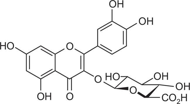
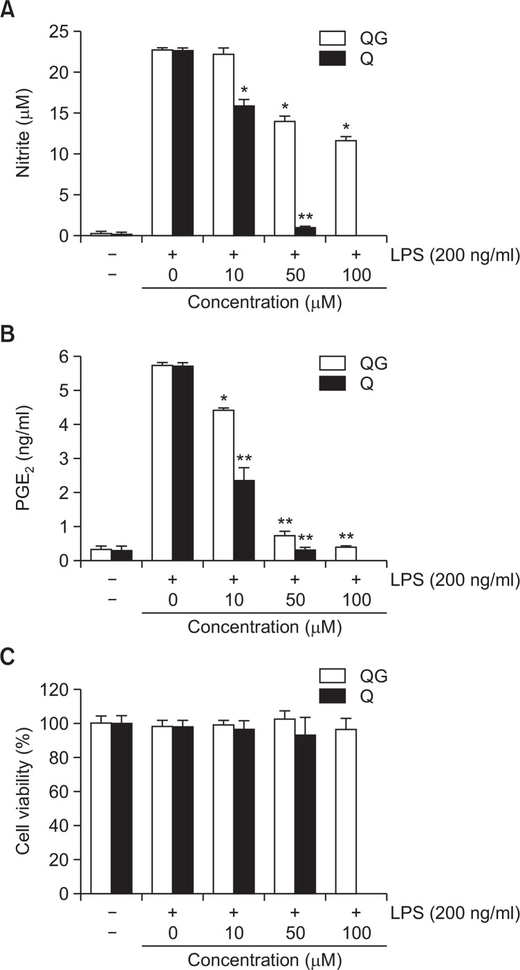
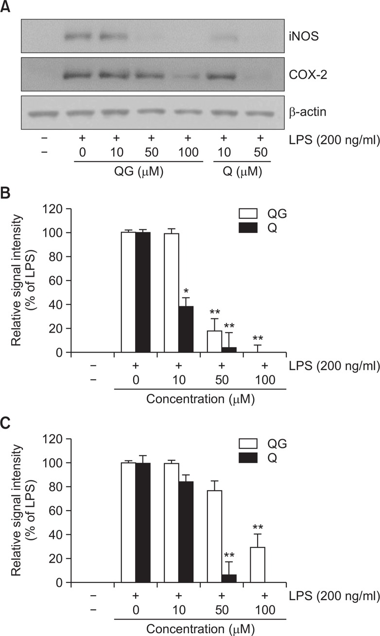
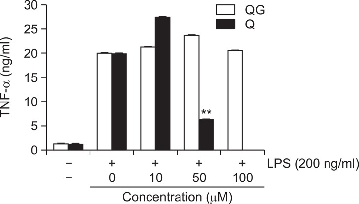
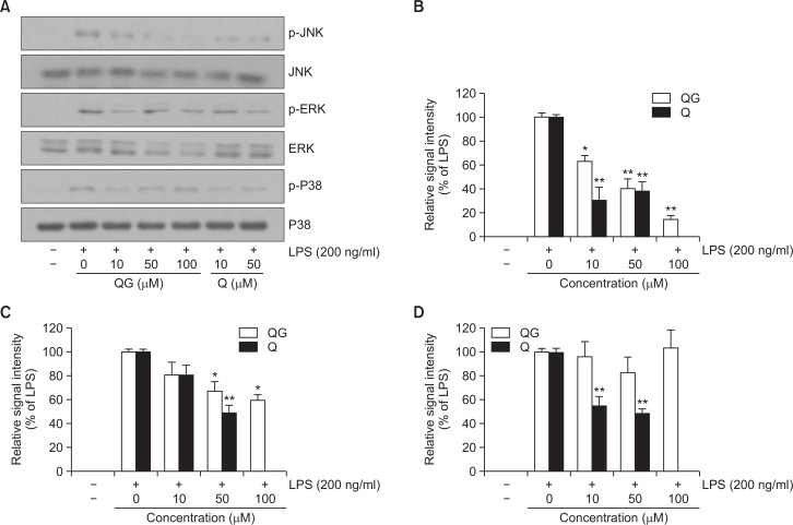
Quercetin, a flavonol, has been reported to exhibit a wide range of biological properties including anti-oxidant and anti-inflammatory activities. However, pharmacological properties of quercetin-3-O-β-D-glucuronide (QG), a glycoside derivative of quercetin, have not been extensively examined. The objective of this study is to elucidate the anti-inflammatory property and underlying mechanism of QG in lipopolysaccharide (LPS)-challenged RAW264.7 macrophage cells in comparison with quercetin. QG significantly suppressed LPS-induced extracellular secretion of pro-inflammatory mediators such as nitric oxide (NO) and PGE₂, and pro-inflammatory protein expressions of iNOS and COX-2. To elucidate the underlying mechanism of the anti-inflammatory property of QG, involvement of MAPK signaling pathways was examined. QG significantly attenuated LPS-induced activation of JNK and ERK in concentration-dependent manners with a negligible effect on p38. In conclusion, the present study demonstrates QG exerts anti-inflammatory activity through the suppression of JNK and ERK signaling pathways in LPS-challenged RAW264.7 macrophage cells.
Keywords: ERK; JNK; Lipopolysaccharide; Quercetin; Quercetin-3-O-β-D-glucuronide; RAW264.7 cells.
Figures
Similar articles
-
Isorhamnetin-3-O-Glucuronide Suppresses JNK and p38 Activation and Increases Heme-Oxygenase-1 in Lipopolysaccharide-Challenged RAW264.7 Cells.Drug Dev Res. 2016 May;77(3):143-51. doi: 10.1002/ddr.21301. Drug Dev Res. 2016. PMID: 27113811
-
N-(p-Coumaryol)-Tryptamine Suppresses the Activation of JNK/c-Jun Signaling Pathway in LPS-Challenged RAW264.7 Cells.Biomol Ther (Seoul). 2014 May;22(3):200-6. doi: 10.4062/biomolther.2014.013. Biomol Ther (Seoul). 2014. PMID: 25009700 Free PMC article.
-
Phosphorylation of Akt Mediates Anti-Inflammatory Activity of 1-p-Coumaroyl β-D-Glucoside Against Lipopolysaccharide-Induced Inflammation in RAW264.7 Cells.Korean J Physiol Pharmacol. 2014 Feb;18(1):79-86. doi: 10.4196/kjpp.2014.18.1.79. Epub 2014 Feb 13. Korean J Physiol Pharmacol. 2014. PMID: 24634601 Free PMC article.
-
Aromadendrin Inhibits Lipopolysaccharide-Induced Nuclear Translocation of NF-κB and Phosphorylation of JNK in RAW 264.7 Macrophage Cells.Biomol Ther (Seoul). 2013 May 30;21(3):216-21. doi: 10.4062/biomolther.2013.023. Biomol Ther (Seoul). 2013. PMID: 24265867 Free PMC article.
-
Quercetin disrupts tyrosine-phosphorylated phosphatidylinositol 3-kinase and myeloid differentiation factor-88 association, and inhibits MAPK/AP-1 and IKK/NF-κB-induced inflammatory mediators production in RAW 264.7 cells.Immunobiology. 2013 Dec;218(12):1452-67. doi: 10.1016/j.imbio.2013.04.019. Epub 2013 May 9. Immunobiology. 2013. PMID: 23735482
Cited by
-
Quercetin-3-O-β-D-glucuronide attenuates osteoarthritis by inhibiting cartilage extracellular matrix degradation and inflammation.J Orthop Translat. 2024 Apr 5;45:236-246. doi: 10.1016/j.jot.2024.01.007. eCollection 2024 Mar. J Orthop Translat. 2024. PMID: 38601200 Free PMC article.
-
Flavonols as potential antiviral drugs targeting SARS-CoV-2 proteases (3CLpro and PLpro), spike protein, RNA-dependent RNA polymerase (RdRp) and angiotensin-converting enzyme II receptor (ACE2).Eur J Pharmacol. 2021 Jan 15;891:173759. doi: 10.1016/j.ejphar.2020.173759. Epub 2020 Nov 27. Eur J Pharmacol. 2021. PMID: 33249077 Free PMC article. Review.
-
Naturally occurring small molecules with dual effect upon inflammatory signaling pathways and endoplasmic reticulum stress response.J Physiol Biochem. 2024 May;80(2):421-437. doi: 10.1007/s13105-024-01014-1. Epub 2024 Mar 19. J Physiol Biochem. 2024. PMID: 38502466 Free PMC article.
-
Rifampicin Alleviates Atopic Dermatitis-Like Response in vivo and in vitro.Biomol Ther (Seoul). 2017 Nov 1;25(6):634-640. doi: 10.4062/biomolther.2017.147. Biomol Ther (Seoul). 2017. PMID: 29081091 Free PMC article.
-
Pharmacological and mechanistic aspects of quercetin in osteoporosis.Front Pharmacol. 2024 Jan 25;15:1338951. doi: 10.3389/fphar.2024.1338951. eCollection 2024. Front Pharmacol. 2024. PMID: 38333006 Free PMC article. Review.
References
-
- Cho SG, Choi EJ. Apoptotic signaling pathways: caspases and stress-activated protein kinases. J Biochem Mol Biol. 2002;35:24–27. - PubMed
-
- Fan D, Zhao Y, Zhou X, Gong X, Zhao C. Simultaneous determination of esculetin, quercetin-3-O-β-D-glucuronide, quercetin-3-O-β-D-glucuronopyranside methyl ester and quercetin in effective part of Polygonum perfoliatum L. using high performace liquid chromatography. Pharmacogn Mag. 2014;10:359–366. doi: 10.4103/0973-1296.137379. - DOI - PMC - PubMed
LinkOut - more resources
Full Text Sources
Other Literature Sources
Research Materials
Miscellaneous
