. 2016 Sep 19;44(16):7722-41.
doi: 10.1093/nar/gkw506. Epub 2016 Jun 2.

The Pch2 AAA+ ATPase promotes phosphorylation of the Hop1 meiotic checkpoint adaptor in response to synaptonemal complex defects

Affiliations

The Pch2 AAA+ ATPase promotes phosphorylation of the Hop1 meiotic checkpoint adaptor in response to synaptonemal complex defects

Esther Herruzo et al. Nucleic Acids Res. .

Abstract

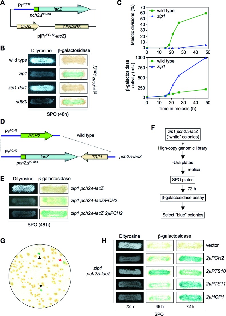
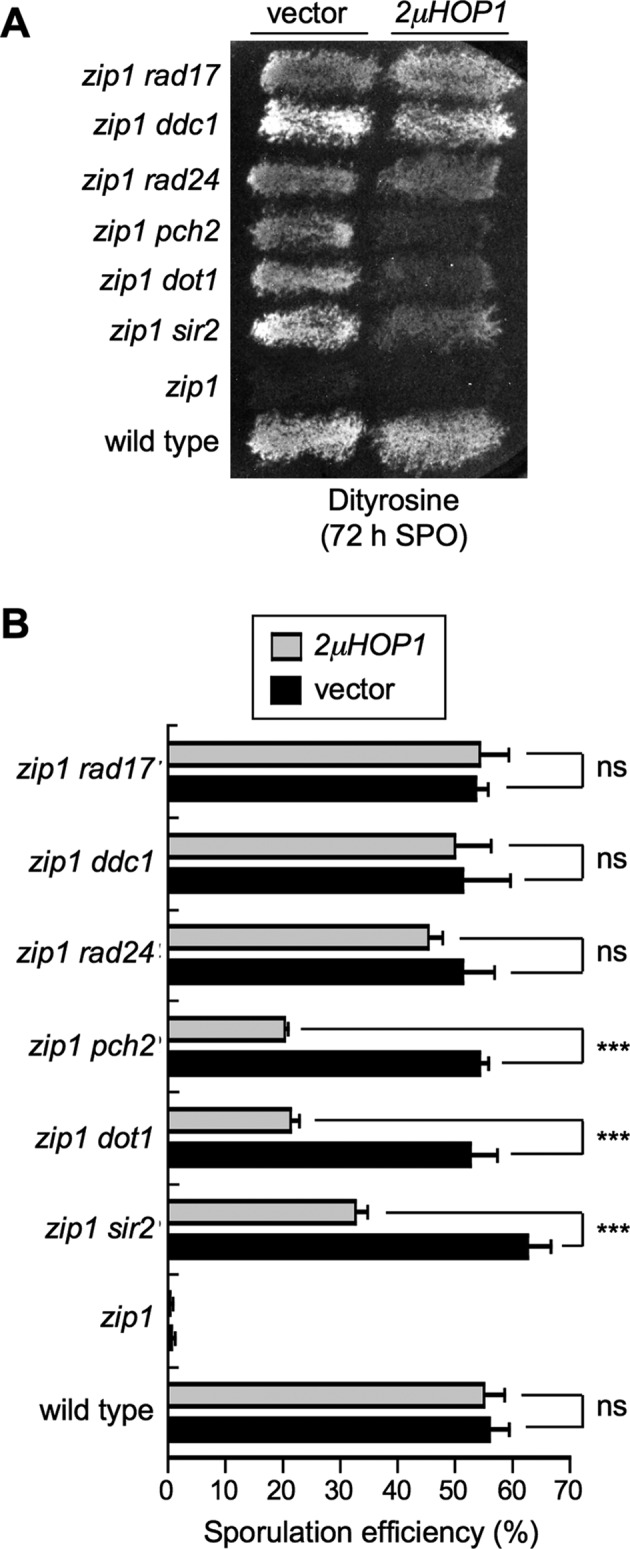
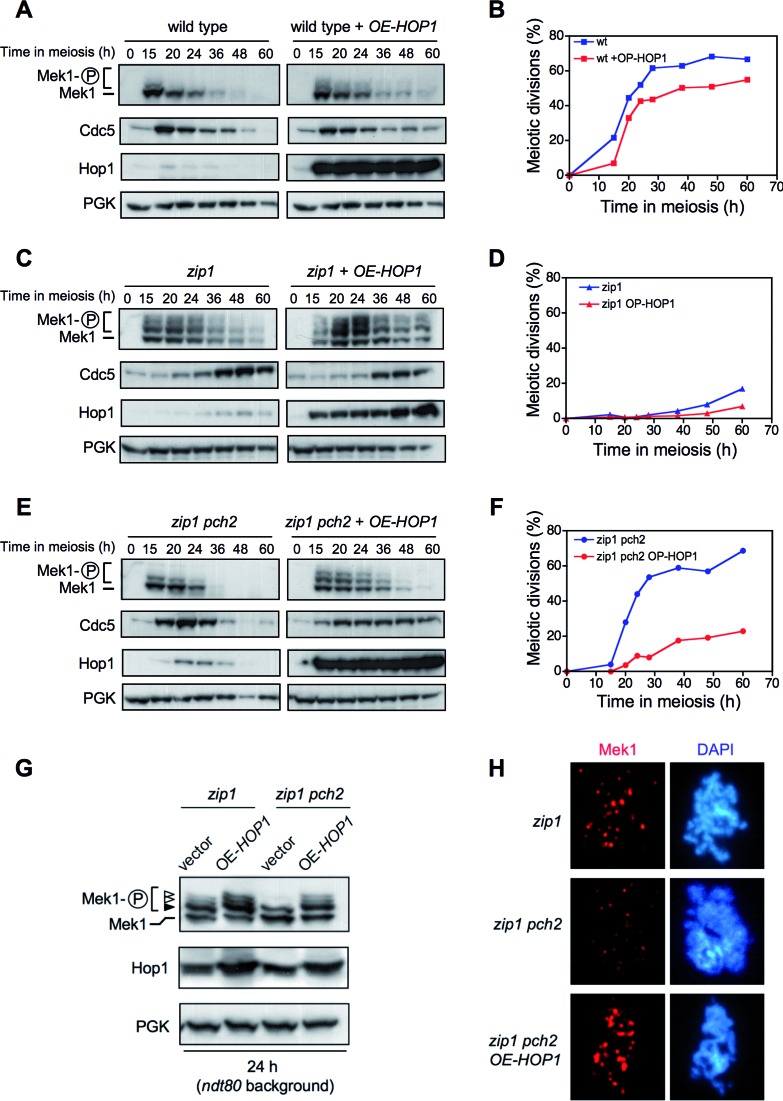
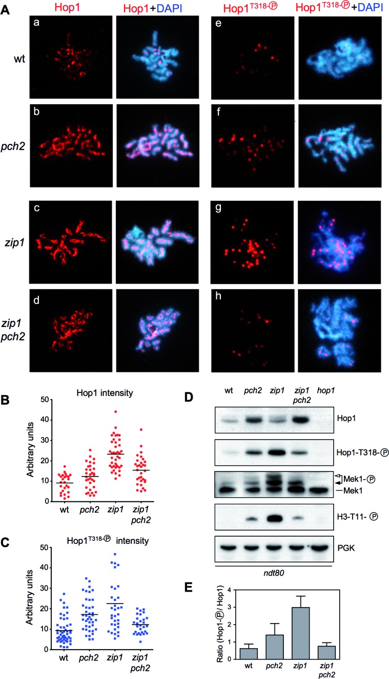
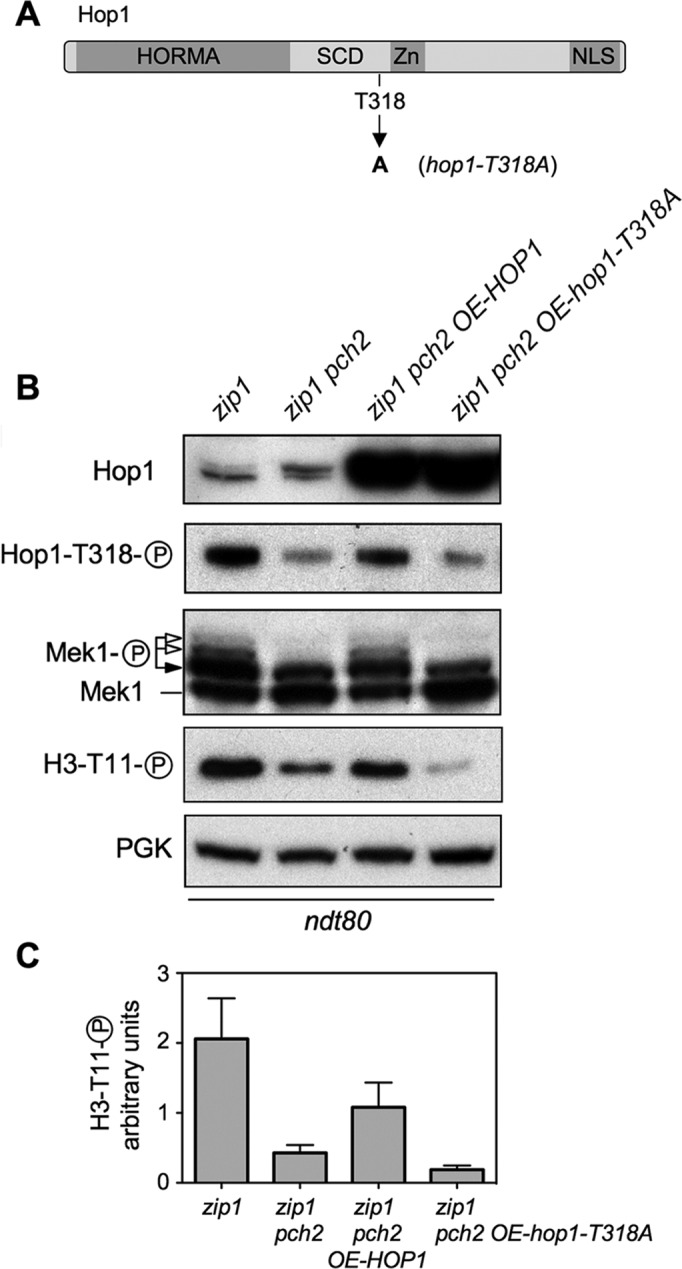
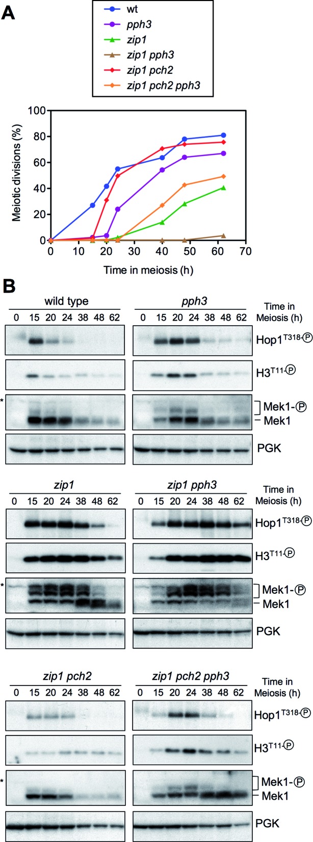
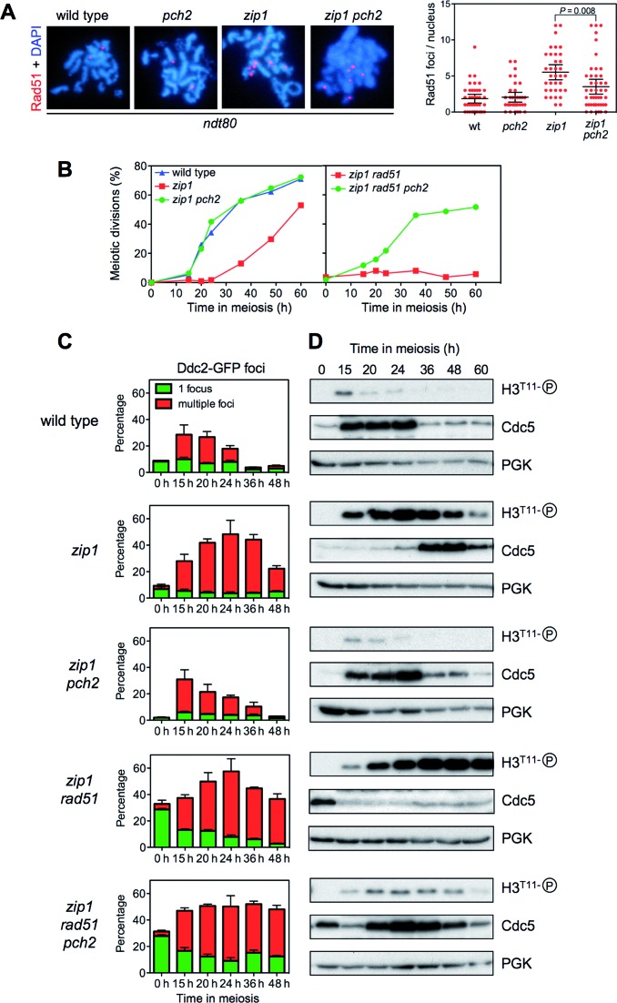
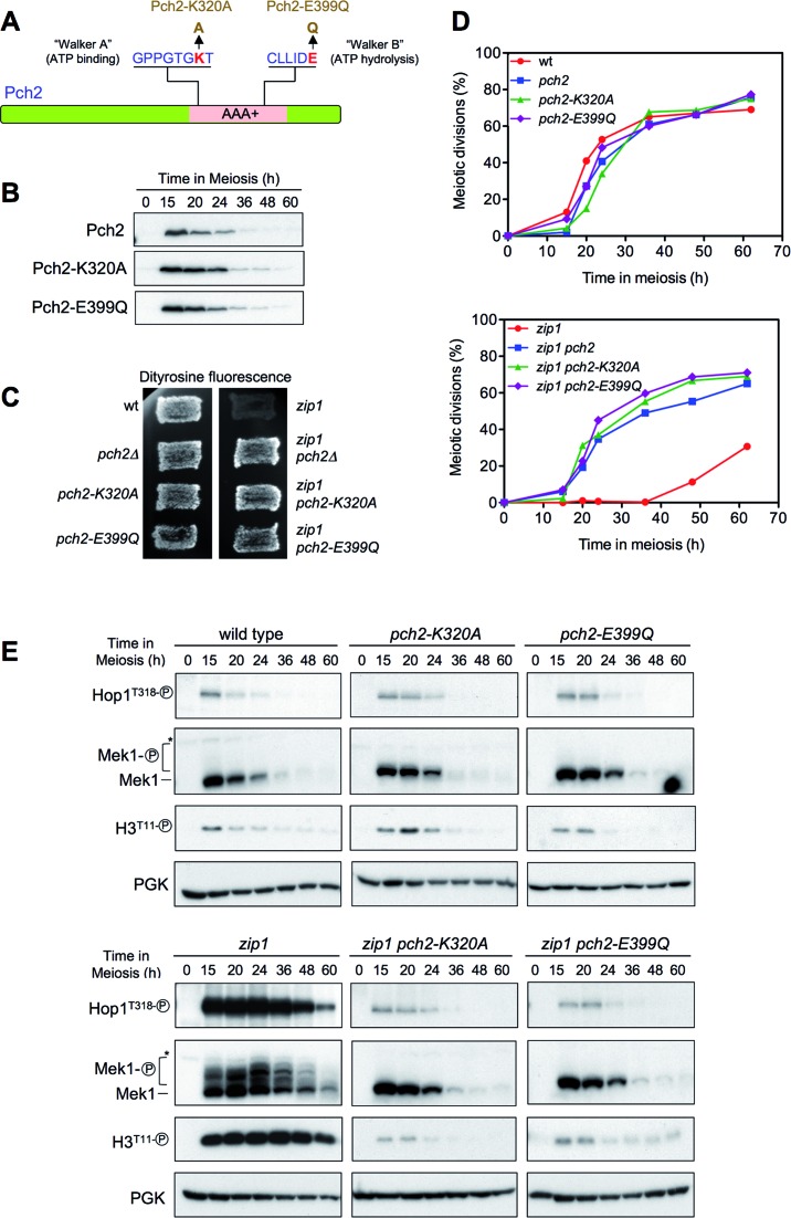
Meiotic cells possess surveillance mechanisms that monitor critical events such as recombination and chromosome synapsis. Meiotic defects resulting from the absence of the synaptonemal complex component Zip1 activate a meiosis-specific checkpoint network resulting in delayed or arrested meiotic progression. Pch2 is an evolutionarily conserved AAA+ ATPase required for the checkpoint-induced meiotic block in the zip1 mutant, where Pch2 is only detectable at the ribosomal DNA array (nucleolus). We describe here that high levels of the Hop1 protein, a checkpoint adaptor that localizes to chromosome axes, suppress the checkpoint defect of a zip1 pch2 mutant restoring Mek1 activity and meiotic cell cycle delay. We demonstrate that the critical role of Pch2 in this synapsis checkpoint is to sustain Mec1-dependent phosphorylation of Hop1 at threonine 318. We also show that the ATPase activity of Pch2 is essential for its checkpoint function and that ATP binding to Pch2 is required for its localization. Previous work has shown that Pch2 negatively regulates Hop1 chromosome abundance during unchallenged meiosis. Based on our results, we propose that, under checkpoint-inducing conditions, Pch2 also possesses a positive action on Hop1 promoting its phosphorylation and its proper distribution on unsynapsed chromosome axes.


Figures

Figure 1.
Figure 1.
Identification of HOP1 in a genetic screen for high-copy suppressors of the zip1 pch2 checkpoint defect using a pch2-lacZ construct as a reporter for meiotic prophase arrest. (A) Schematic representation of a centromeric plasmid (pSS51) carrying the PCH2 promoter and the coding sequence for the first N-terminal 90 amino acids fused in frame with the bacterial lacZ gene. (B) Dityrosine fluorescence and β-galactosidase assays of the indicated strains transformed with the pSS51 plasmid after 48 h on sporulation plates. (C) Kinetics of meiotic divisions and β-galactosidase activity in meiotic time courses of wild-type and zip1 strains containing the pSS51 plasmid. (D) Schematic representation of the substitution of the PCH2 gene for the pch2-lacZ construct at the genomic locus. (E) Dityrosine fluorescence and β-galactosidase assays of the indicated strains after 48 h on sporulation plates. (F) Scheme of the genetic screen. (G) Representative β-galactosidase assay of a plate from zip1 pch2Δ-lacZ transformed with the genomic high-copy library. Black triangles indicate positive controls deliberately placed at known positions on every plate of the screen. The red arrow points to a positive ‘blue’ candidate. (H) Dityrosine fluorescence and β-galactosidase assays of the zip1 pch2Δ-lacZ strain transformed with the indicated plasmids. Strains for (B) and (C) are: BR2495 (wild type), MY63 (zip1), DP174 (zip1 dot1) and S3483 (ndt80). Strains for (E), (F), (G) and (H) are: DP221 (zip1 pch2Δ-lacZ) and DP228 (zip1 pch2Δ-lacZ/PCH2). DP221 was transformed with empty vector or the indicated high-copy plasmids.
Figure 2.
Figure 2.
High-copy HOP1 specifically suppresses the checkpoint defect of zip1 pch2, zip1 sir2 and zip1 dot1. (A) Dityrosine fluorescence assay of the indicated strains transformed with empty vector (YEp352) of high-copy HOP1 (R1692) after 3 days on sporulation plates. (B) Microscopic quantification of the sporulation efficiency of the strains analyzed in (A). Three independent counts were performed. Means and standard deviations are shown. (ns), no significant difference; (***), P < 0.001. Strains are: BR2495 (wild type), MY63 (zip1), DP174 (zip1 dot1), DP223 (zip1 pch2), DP267 (zip1 sir2), S4295 (zip1 rad24), S4278 (zip1 ddc1) and S4286 (zip1 rad17).
Figure 3.
Figure 3.
HOP1 overexpression largely restores meiotic arrest, Mek1 activation, Mek1 localization and delayed Cdc5 production in zip1 pch2. Western blot analysis of the indicated proteins and meiotic kinetics of (A and B) wild type, (C and D) zip1 and (E and F) zip1 pch2 transformed with empty vector (pRS426) or high-copy HOP1 (R1692). Strains are: DP421 (wild type), DP422 (zip1) and DP1029 (zip1 pch2). (G) Analysis of Mek1 phosphorylated forms in ndt80-arrested strains. The black arrowhead marks the Mec1/Tel1-dependent band and the white arrowheads point to the forms resulting from Mek1 autophosphorylation (8). Strains are: DP428 (zip1) and DP881 (zip1 pch2), transformed with pRS426 (empty vector) or R1692 (OE-HOP1). (H) Analysis of Mek1 localization by immunofluorescence of spread meiotic chromosomes using anti-GFP antibodies. Representative nuclei are shown. Strains are DP582 (zip1) and DP1111 (zip1 pch2).
Figure 4.
Figure 4.
Pch2 promotes Hop1T318 phosphorylation in zip1. (A) Immunofluorescence of spread meiotic chromosomes stained with DAPI (blue) and anti-Hop1 (a-d panels) or anti-phospho-Hop1T318 (e–h panels) antibodies (red). Representative nuclei are shown. (B and C) Quantification of total Hop1 and phospho-Hop1T318 fluorescence signal, respectively, on the spreads analyzed in (A). Each spot in the plot represents the intensity of a nucleus scored. (D) Western blot analysis of the indicated proteins and phosphorylation events. (E) Quantification of relative Hop1T318 phosphorylation analyzed as in (D). The ratio of phospho-Hop1T318 versus total Hop1 chemiluminiscence signal is represented. Means and standard deviations from three independent experiments are shown. Strains are: DP424 (wild type), DP1058 (pch2), DP428 (zip1), DP881 (zip1 pch2) and DP700 (hop1). Spreads and lysates were prepared 24 h after meiotic induction of ndt80 cells. For DP700 the sample was taken at 17 h.
Figure 5.
Figure 5.
Hop1T318 phosphorylation is the critical checkpoint event impaired in zip1 pch2. (A) Schematic representation of the Hop1 protein domains and the position of the mutated T318 phosphosite (6) (B) Overexpression of wild-type HOP1, but not the hop1-T318A mutant, restores Mek1 activation in zip1 pch2. Western blot analysis of Hop1, phospho-Hop1T318, Mek1 and phospho-H3T11 in ndt80-arrested cells. (C) Quantification of the phospho-H3T11 signal from three experiments. Note that Mek1 activity markedly increases, but is not fully restored in zip1 pch2 OE-HOP1 cells due to plasmid-loss events in the meiotic cultures (43); those cells that lose the plasmid do not overproduce Hop1 and do not contribute to Mek1 phosphorylation. Strains are DP428 (zip1) and DP881 (zip1 pch2) transformed with pRS426 (empty vector), pSS316 (OE-HOP1) or pSS317 (OE-hop1-T318A).
Figure 6.
Figure 6.
Impact of the PP4 phosphatase on Pch2-dependent meiotic checkpoint. (A) Time course of meiotic nuclear divisions; the percentage of cells containing two or more nuclei is represented. (B) Western blot analysis of Hop1T318 phosphorylation and Mek1 activity at the indicated time points in meiosis. PGK was used as a loading control. Strains are: DP421 (wild type), DP1247 (pph3), DP422 (zip1), DP1249 (zip1 pph3), DP1029 (zip1 pch2) and DP1245 (zip1 pch2 pph3).
Figure 7.
Figure 7.
Effect of Pch2 on cell cycle progression and resolution of zip1-induced recombination intermediates. (A) Localization and quantification of Rad51 foci as markers for unrepaired DSBs on spread meiotic nuclei of ndt80 cells after 24 h of meiotic induction. Strains are: DP424 (wild type), DP1058 (pch2), DP428 (zip1) and DP881 (zip1 pch2). (B) Time course of meiotic nuclear divisions. The percentage of cells with two or more nuclear masses is represented. (C) Quantification of Ddc2-GFP foci throughout meiosis. The percentage of cells containing a single non-meiotic Ddc2 focus (green bars) or multiple meiotic Ddc2 foci (red bars) from three different counts is represented. Note that rad51 cells accumulate spontaneous non-meiotic Ddc2 foci at time zero. Between 150 and 800 cells were scored for each strain at every time point. (D) Western blot analysis of phospho-H3T11 (as a reporter for Mek1 activity), Cdc5 (as a marker for meiosis I entry) and PGK (as a loading control) from the same cultures analyzed in (B) and (C). Strains for (B), (C) and (D) are: DP448 (wild type), DP449 (zip1), DP1379 (zip1 pch2) DP1381 (zip1 rad51) and DP1382 (zip1 pch2 rad51).
Figure 8.
Figure 8.
The ATPase activity of Pch2 is required for its checkpoint function. (A) Schematic representation of the Pch2 protein indicating the AAA+ domain, the conserved Walker A and Walker B motifs, and the mutations introduced at both sites. (B) Western blot analysis of Pch2, Pch2-K320A or Pch2-E399Q production (detected with anti-HA antibodies) during meiosis. (C) Dityrosine fluorescence assay. (D) Time course of meiotic nuclear divisions; the percentage of cells containing two or more nuclei is represented. (E) Western blot analysis of Hop1T318 phosphorylation and Mek1 activation. PGK was used as a loading control. Strains are: DP1151 (wild type), DP1164 (pch2Δ), DP1163 (pch2-K320A), DP1287 (pch2-E399Q), DP1152 (zip1), DP1161 (zip1 pch2Δ), DP1162 (zip1 pch2-K320A) and DP1288 (zip1 pch2-E399Q).
Figure 9.
Figure 9.
Localization of ATPase-deficient versions of Pch2. (A) Immunofluorescence of meiotic chromosomes stained with anti-HA or anti-MYC antibodies to detect Pch2, Pch2-K320A or Pch2-E399Q (red), anti-Hop1 antibodies (green) and DAPI (blue). Strains are: DP1243 (wild type), DP1193 (pch2-K320A), DP1262 (pch2-E399Q), DP1244 (zip1), DP1192 (zip1 pch2-K320A) and DP1263 (zip1 pch2-E399Q). (B) Immunofluorescence of whole meiotic cells stained with anti-HA antibodies (to detect Pch2 or Pch2-K320A; red), anti-Hop1 antibodies (green) and DAPI (blue). The contour of the cells is outlined in the rightmost panels. Representative cells 24 h after meiotic induction in zip1 ndt80 background are shown. The arrows point to the rDNA region, which is distinguishable by the accumulation of Pch2 and the absence of Hop1. This region is not recognizable in the pch2-K320A mutant due to the mislocalization of Pch2-K320A and Hop1. Strains are: DP1190 (wild type) and DP1192 (pch2-K320A).
Figure 10.
Figure 10.
Mutation of the Pch2 ATP-binding site impairs the stability of the hexameric complex. (A) Whole cell extracts (WCE) prepared after 16 h in meiosis were immunoprecipitated with anti-HA antibodies. WCE and immunoprecipitates (IP) were analyzed by Western blot with both anti-HA and anti-Myc antibodies, as indicated. Strains are: DP1325 (PCH2-HA/PCH2-Myc; lane 1), DP1329 (PCH2/PCH2-Myc; lane 2) and DP1337 (pch2-K320A-HA/pch2-K320A-Myc; lane 3). To discard a problem with detection levels, in lane 2 (HA-untagged control) and lane 3, three more times of IP compared to lane 1 were loaded. (B) Schematic interpretation of the result described in (A). K and A represent a lysine and an alanine, respectively, at position 320 of Pch2.
Figure 11.
Figure 11.
A model for Pch2 checkpoint function. (A) In unperturbed wild-type meiosis, nucleolar Pch2 excludes Hop1 from the rDNA preventing recombination at this chromosome XII array. In turn, chromosomal Pch2 dictates Hop1 discontinuous axis distribution sustaining proper synapsis and recombination. In the pch2 single mutant, Hop1 localizes to the nucleolar region and unwanted rDNA recombination occurs. Hop1 is also more abundant on chromosome axes disturbing normal recombination events that slightly increase Hop1-T318 phosphorylation. Nevertheless, most recombination defects of the pch2 mutant are only evident when DSBs are limiting (28,56). (B) In the synapsis-deficient zip1 mutant, Pch2 is absent from the chromosomes and concentrated in the rDNA. This configuration supports continuous distribution of Hop1 along unsynapsed axes and high levels of Hop1-T318 phosphorylation relaying the checkpoint signal to Mek1 activation. When PCH2 is deleted (or the ATPase inactivated), Hop1 loading and phosphorylation is impaired leading to inoperative checkpoint. (C) Despite the weakened Hop1/Mek1 activation, HOP1 overexpression in zip1 pch2 provides enough protein to restore high levels of global Hop1 phosphorylation and reinstate checkpoint function (see text for additional details).

Similar articles

Cited by

References

    1. MacQueen A.J., Hochwagen A. Checkpoint mechanisms: the puppet masters of meiotic prophase. Trends Cell. Biol. 2011;21:393–400. - PubMed
    1. Roeder G.S., Bailis J.M. The pachytene checkpoint. Trends Genet. 2000;16:395–403. - PubMed
    1. Subramanian V.V., Hochwagen A. The meiotic checkpoint network: step-by-step through Meiotic Prophase. Cold Spring Harb. Perspect. Biol. 2014;6:e1002369. - PMC - PubMed
    1. Schwacha A., Kleckner N. Interhomolog bias during meiotic recombination: meiotic functions promote a highly differentiated interhomolog-only pathway. Cell. 1997;90:1123–1135. - PubMed
    1. Sym M., Engebrecht J.A., Roeder G.S. ZIP1 is a synaptonemal complex protein required for meiotic chromosome synapsis. Cell. 1993;72:365–378. - PubMed

MeSH terms