

Absolute number of parvicellular and magnocellular neurons in the red nucleus of the rat midbrain: a stereological study
- PMID: 27257130
- PMCID: PMC4974543
- DOI: 10.1111/joa.12495
Absolute number of parvicellular and magnocellular neurons in the red nucleus of the rat midbrain: a stereological study
Abstract
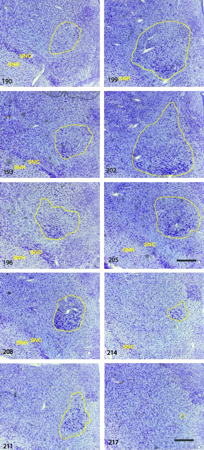
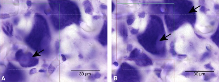
The absolute number of parvicellular and magnocellular neurons in the red nucleus was estimated using design-based stereological counting methods and systematic random sampling techniques. Six young adult male rats, and a complete set of serial 40-μm glycolmethacrylate sections for each rat, were used to quantify neuronal numbers. After a random start, a systematic subset (i.e. every third) of the serial sections was used to estimate the total volume of the red nucleus using Cavalieri's method. The same set of sampled sections was used to estimate the number of neurons in a known subvolume (i.e. the numerical density Nv ) by the optical disector method. Multiplication of the total volume by Nv yielded the absolute number of neurons. It was found that the right red nucleus consisted, on average, of 8400 parvicellular neurons (with a coefficient of variation of 0.16) and 7000 magnocellular neurons (0.12). These total neuronal numbers provide important data for the transfer of information through these nuclei and for species comparisons.
Keywords: Cavalieri's method; magnocellular neurons; optical disector method; parvicellular neurons; red nucleus; species comparisons; transfer of information.
© 2016 Anatomical Society.
Figures
Similar articles
-
Total number of neurons in the habenular nuclei of the rat epithalamus: a stereological study.J Anat. 2006 May;208(5):577-85. doi: 10.1111/j.1469-7580.2006.00573.x. J Anat. 2006. PMID: 16637880 Free PMC article.
-
Total number of neurons in the neostriatal, pallidal, subthalamic, and substantia nigral nuclei of the rat basal ganglia: a stereological study using the cavalieri and optical disector methods.J Comp Neurol. 1996 Mar 18;366(4):580-99. doi: 10.1002/(SICI)1096-9861(19960318)366:4<580::AID-CNE3>3.0.CO;2-0. J Comp Neurol. 1996. PMID: 8833111
-
Analysis of cell death in the trochlear nucleus of the chick embryo: calibration of the optical disector counting method reveals systematic bias.J Comp Neurol. 1999 Jun 28;409(2):169-86. J Comp Neurol. 1999. PMID: 10379913
-
A brief update on physical and optical disector applications and sectioning-staining methods in neuroscience.J Chem Neuroanat. 2018 Nov;93:16-29. doi: 10.1016/j.jchemneu.2018.02.009. Epub 2018 Feb 26. J Chem Neuroanat. 2018. PMID: 29496551 Review.
-
Stereological methods for estimating the total number of neurons and synapses: issues of precision and bias.Trends Neurosci. 1999 Feb;22(2):51-61. doi: 10.1016/s0166-2236(98)01362-9. Trends Neurosci. 1999. PMID: 10092043 Review.
Cited by
-
Delayed Double Treatment with Adult-Sourced Adipose-Derived Mesenchymal Stem Cells Increases Striatal Medium-Spiny Neuronal Number, Decreases Striatal Microglial Number, and Has No Subventricular Proliferative Effect, after Acute Neonatal Hypoxia-Ischemia in Male Rats.Int J Mol Sci. 2021 Jul 23;22(15):7862. doi: 10.3390/ijms22157862. Int J Mol Sci. 2021. PMID: 34360638 Free PMC article.
-
Systematic Methods for Isolating High Purity Nuclei from Ten Important Plants for Omics Interrogation.Cells. 2022 Dec 3;11(23):3919. doi: 10.3390/cells11233919. Cells. 2022. PMID: 36497177 Free PMC article.
-
Neural Signals in Red Nucleus during Reactive and Proactive Adjustments in Behavior.J Neurosci. 2020 Jun 10;40(24):4715-4726. doi: 10.1523/JNEUROSCI.2775-19.2020. Epub 2020 May 6. J Neurosci. 2020. PMID: 32376779 Free PMC article.
-
The diversity and plasticity of descending motor pathways rewired after stroke and trauma in rodents.Front Neural Circuits. 2025 Mar 21;19:1566562. doi: 10.3389/fncir.2025.1566562. eCollection 2025. Front Neural Circuits. 2025. PMID: 40191711 Free PMC article. Review.
References
-
- Bjugn R, Gundersen HJG (1993) Estimate of the total number of neurons and glial and endothelial cells in the rat spinal cord by means of the optical disector. J Comp Neurol 328, 406–414. - PubMed
-
- Boseila AWA, Hashem SM, Badawy YH (1975) Volumetric studies on the red nucleus of the rat at different ages. Acta Anat 91, 175–180. - PubMed
-
- Boyce RW, Dorph‐Petersen K‐A, Lyck L, et al. (2010) Design‐based stereology: introduction to basic concepts and practical approaches for estimation of cell number. Toxicol Pathol 38, 1011–1025. - PubMed
-
- Braendgaard H, Evans SM, Howard CV, et al. (1990) Total number of neurons in the human neocortex unbiasedly estimated using optical disectors. J Microsc 157, 285–304. - PubMed
-
- Cameron SH, Alwakeel A, Goddard L, et al. (2015) Delayed post‐treatment with bone marrow‐derived mesenchymal stem cells is neurorestorative of striatal medium‐spiny neurons and improves motor function after neonatal rat hypoxia‐ischemia. Mol Cell Neurosci 68, 56–72. - PubMed
MeSH terms
LinkOut - more resources
Full Text Sources
Other Literature Sources
