. 2016 Jun 3:6:27060.
doi: 10.1038/srep27060.

Unexpected involvement of staple leads to redesign of selective bicyclic peptide inhibitor of Grb7

Affiliations

Unexpected involvement of staple leads to redesign of selective bicyclic peptide inhibitor of Grb7

Menachem J Gunzburg et al. Sci Rep. .

Abstract

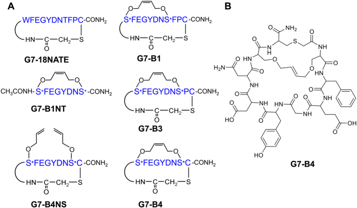
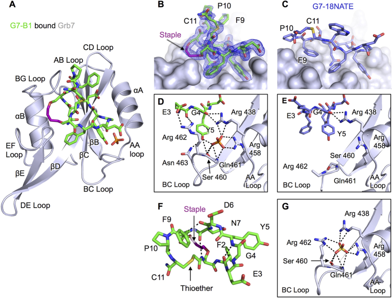
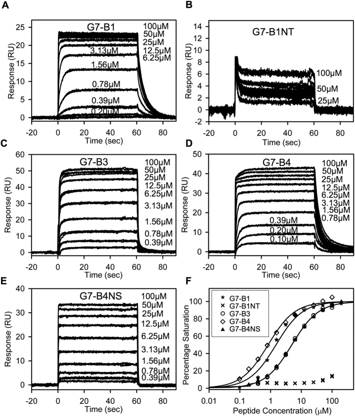
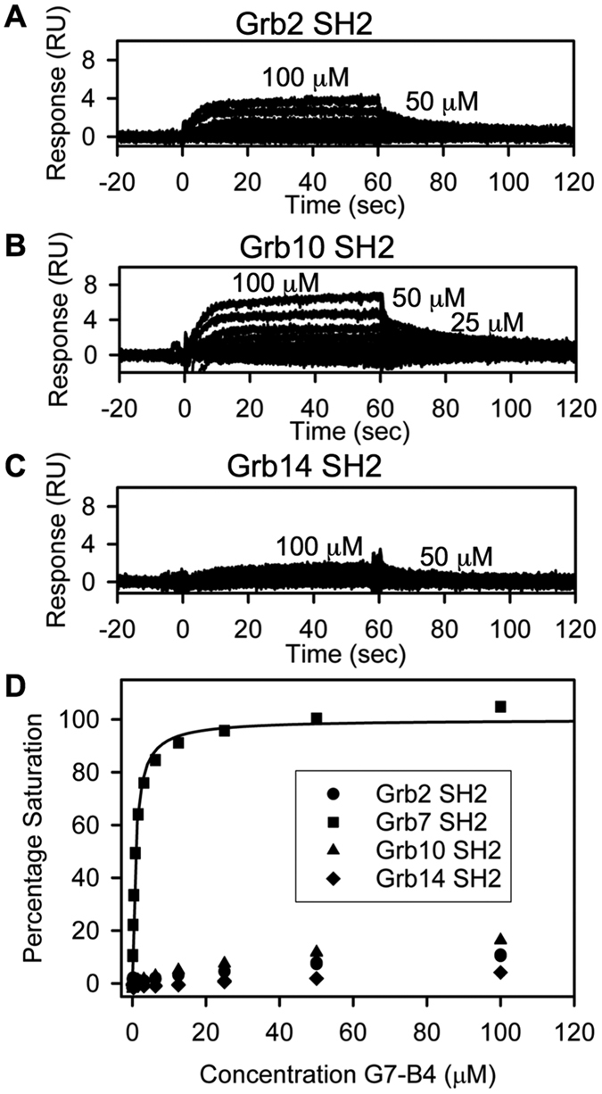
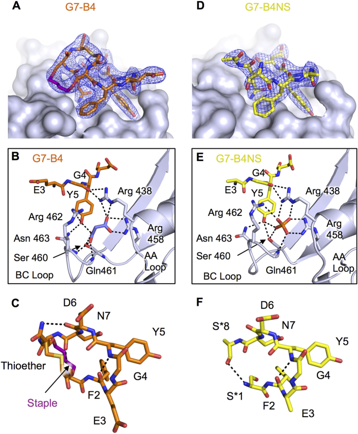
The design of potent and specific peptide inhibitors to therapeutic targets is of enormous utility for both proof-of-concept studies and for the development of potential new therapeutics. Grb7 is a key signaling molecule in the progression of HER2 positive and triple negative breast cancers. Here we report the crystal structure of a stapled bicyclic peptide inhibitor G7-B1 in complex with the Grb7-SH2 domain. This revealed an unexpected binding mode of the peptide, in which the staple forms an alternative contact with the surface of the target protein. Based on this structural information, we designed a new series of bicyclic G7 peptides that progressively constrain the starting peptide, to arrive at the G7-B4 peptide that binds with an approximately 2-fold enhanced affinity to the Grb7-SH2 domain (KD = 0.83 μM) compared to G7-B1 and shows low affinity binding to Grb2-, Grb10- and Grb14-SH2 domains (KD > 100 μM). Furthermore, we determined the structure of the G7-B4 bicyclic peptide in complex with the Grb7-SH2 domain, both before and after ring closing metathesis to show that the closed staple is essential to the target interaction. The G7-B4 peptide represents an advance in the development of Grb7 inhibitors and is a classical example of structure aided inhibitor development.


Figures

Figure 1
Figure 1. Schematic showing the structures of the 6 peptides referred to in this paper.
(A) Schematic depicts amino acid single letter code in blue, with S* referring to O-allylserine. Other chemical functionalities are depicted in black. (B) chemical structure diagram of the G7-B4 peptide.
Figure 2
Figure 2. Structure of the G7-B1 and Grb7-SH2 domain complex in comparison to other Grb7-SH2 structures.
(A) Cartoon representation of the Grb7-SH2 domain (grey), with G7-B1 (green) and phosphate ion in stick representation. (B) G7-B1 peptide shown in green stick representation at the surface of Grb7-SH2 domain (grey), with 2Fo-Fc map surrounding G7-B1 shown contoured at 1.2σ. (C) G7-18NATE (purple sticks) bound to Grb7-SH2 domain (grey surface) (PDB ID: 3PQZ) in the same orientation as in (B). (D) G7-B1 (green sticks) oriented to show intramolecular hydrogen bond and electrostatic interactions at the pY binding pocket of Grb7-SH2 (grey cartoon). (E) Grb7-SH2 domain (grey cartoon) pY binding pocket interactions with G7-18NATE (purple sticks) (PDB ID: 3PQZ) in the same orientation as in (D). (E) G7-B1 structure (green sticks) shown in absence of Grb7-SH2 binding partner to highlight intramolecular hydrogen bonds. (G) apo-Grb7-SH2 domain pY binding pocket (grey cartoon) showing bound sulfate (PDB ID: 2QMS).
Figure 3
Figure 3. Binding affinity determination of G7 peptides for Grb7-SH2.
(A–F) SPR Sensorgrams for G7 peptides (0–100 μM) binding to the Grb7-SH2 domain. The peptide samples were injected from 0 to 60 s; otherwise, buffer was flowing. (A) G7-B1. (B) G7-B1NT. Labels for low peptide concentrations are omitted for clarity. (C) G7-B3. (D) G7-B4. (E) G7-B4NS. (F) Equilibrium binding curves for peptides binding to the Grb7-SH2 domain (shown using a logarithmic scale for clarity). Percentage saturation for G7-B1NT was calculated as Bmax of 0.64 × Theroretical Bmax (based on the average Bmax/Theroretical Bmax ratio for all other peptides). Fits to a single-site binding model are shown as solid lines. Data for G7-B4 served as a control in Fig. 4D.
Figure 4
Figure 4. Determination of binding specificity of G7-B4 for Grb7-SH2.
(A–C) Sensorgrams for G7-B4 (0–100 μM) binding to different Grb protein SH2 domains. The G7-B4 samples were injected from 0 to 60 s; otherwise, buffer was flowing. Labels for low G7-B4 concentrations are omitted for clarity. (A) Binding to Grb2-SH2 domain. (B) Binding to Grb10-SH2 domain. (D) Binding to Grb14-SH2 domain. (D) Equilibrium binding curves for G7-B4 binding to different Grb SH2 domains. Percentage saturation for Grb2-SH2, Grb10 SH2 and Grb14 SH2 were calculated as Bmax of 0.64 × Theroretical Bmax (based on the average Bmax/Theroretical Bmax ratio for peptides binding Grb7). Fit to a single-site binding model is shown as solid line. Data for G7-B4 binding to Grb7-SH2 was also used in Fig. 3F.
Figure 5
Figure 5. Structure of the G7-B4 and G7-B4NS peptide in complex with Grb7-SH2.
(A) G7-B4 peptide shown in orange stick representation at the surface of Grb7-SH2 domain (grey), with 2Fo-Fc map surrounding G7-B4 shown contoured at 1.2σ. (B) Details of the G7-B4 bound pY binding pocket. G7-B4 and malonic acid (purple sticks) bound to Grb7-SH2. Key amino acids are shown as sticks and hydrogen bonds as dashed lines. (C) G7-B4 oriented to show intramolecular hydrogen bonds. (D) G7-B4NS peptide shown in yellow stick representation at the surface of Grb7-SH2 domain (grey), with 2Fo-Fc map surrounding G7-B4NS shown contoured at 1.2σ. (E) Details of the G7-B4NS pY binding pocket. G7-B4NS and phosphate bound to Grb7-SH2. Key amino acids are shown as sticks and hydrogen bonds as dashed lines. (F) G7-B4NS oriented to show intramolecular hydrogen bonds.

References

    1. Zinzalla G. & Thurston D. E. Targeting protein-protein interactions for therapeutic intervention: a challenge for the future. Future Med Chem 1, 65–93 (2009). - PubMed
    1. Benyamini H. & Friedler A. Using peptides to study protein-protein interactions. Future Med Chem 2, 989–1003 (2010). - PubMed
    1. Maes M., Loyter A. & Friedler A. Peptides that inhibit HIV-1 integrase by blocking its protein-protein interactions. FEBS J 279, 2795–2809 (2012). - PubMed
    1. Gao M., Cheng K. & Yin H. Targeting protein-protein interfaces using macrocyclic peptides. Biopolymers 104, 310–316 (2015). - PMC - PubMed
    1. Jubb H., Blundell T. L. & Ascher D. B. Flexibility and small pockets at protein-protein interfaces: New insights into druggability. Prog. in Biophysics and Molecular Biology 119, 2–9 (2015). - PMC - PubMed

Publication types