. 2016 Jun 1;30(11):1278-88.
doi: 10.1101/gad.280495.116. Epub 2016 Jun 2.

Jmjd2/Kdm4 demethylases are required for expression of Il3ra and survival of acute myeloid leukemia cells

Affiliations

Jmjd2/Kdm4 demethylases are required for expression of Il3ra and survival of acute myeloid leukemia cells

Karl Agger et al. Genes Dev. .

Abstract

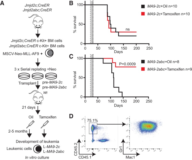
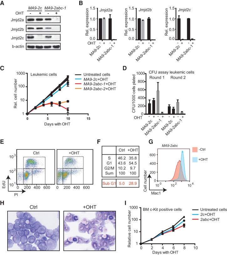
Acute myeloid leukemias (AMLs) with a rearrangement of the mixed-linage leukemia (MLL) gene are aggressive hematopoietic malignancies. Here, we explored the feasibility of using the H3K9- and H3K36-specific demethylases Jmjd2/Kdm4 as putative drug targets in MLL-AF9 translocated leukemia. Using Jmjd2a, Jmjd2b, and Jmjd2c conditional triple-knockout mice, we show that Jmjd2/Kdm4 activities are required for MLL-AF9 translocated AML in vivo and in vitro. We demonstrate that expression of the interleukin 3 receptor α (Il3ra also known as Cd123) subunit is dependent on Jmjd2/Kdm4 through a mechanism involving removal of H3K9me3 from the promoter of the Il3ra gene. Importantly, ectopic expression of Il3ra in Jmjd2/Kdm4 knockout cells alleviates the requirement of Jmjd2/Kdm4 for the survival of AML cells, showing that Il3ra is a critical downstream target of Jmjd2/Kdm4 in leukemia. These results suggest that the JMJD2/KDM4 proteins are promising drug targets for the treatment of AML.

Keywords: H3K9 methylation; JMJD2; acute myeloid leukemia; epigenetics; histone demethylase; interleukin 3.


Figures

Figure 1.
Figure 1.
MLL-AF9 cells are dependent on the combined activity of Jmjd2a, Jmjd2c, and Jmjd2c in vivo. (A) Schematic drawing of the experimental setup. (B,C) Kaplan-Meier curve depicting the survival of mice transplanted with 5 × 10⁴ preleukemic cells with the indicated genotypes. Twenty-one days after transplantation, the mice were injected daily with 1 mg of tamoxifen dissolved in oil or with oil alone for a period of 10 d (indicated as a gray area in the growth curve). The median survival of MA9-2abc mice injected with oil was 112.5 d; for MA9-2abc mice injected with tamoxifen, the median survival was not reached; for MA9-2c mice injected with oil, the median survival was 99.5 d; and for MA9-2c mice injected with tamoxifen, the median survival was 93 d. P-values are the result of a Mantel-Cox statistical test. (D) Representative FACS plots of cells from the spleen of a leukemic mouse. Plots show Gr1+ and Mac1+ double-positive in the CD45.2 gate.
Figure 2.
Figure 2.
Loss of Jmjd2/Kdm4 activity results in decreased growth combined with an increase in differentiation and cell death for MLL-AF9 transformed leukemic cells. (A) Western blot documenting efficient depletion of Jmjd2a, Jmjd2b, and Jmjd2c protein levels in MLL-AF9 transformed murine leukemic cells after 96 h of treatment with 500 nM OHT. (B) RT-qPCR from the indicated cells, confirming efficient depletion of full-length Jmjd2a, Jmjd2b, and Jmjd2c mRNA after OHT treatment. (C) Growth curve of three different leukemic cell lines (L-MA9-2c, L-MA9-2abc-1, and L-MA9-2abc-2) in liquid culture with or without 500 nM OHT. The cell lines were established from leukemic mice transplanted with preleukemic cells of the indicated genotypes. (D) Blast colony formation assay of MA9-2c and MA9-2abc leukemic cells in methylcellulose. Cells were plated after 4 d of treatment with 500 nM OHT in liquid medium. (E) FACS analysis of EdU incorporation in MA9-2abc cells showing an increased sub-G1 fraction, indicative of cell death. (F) The top panel represents cell cycle distribution of MA9-2abc cells, excluding the sub-G1 fraction. The bottom red panel shows the percentage of cells with a sub-G1 (<2N) DNA content. The cells were treated for 96 h with 500 nM OHT prior to the analysis. (G) FACS plot showing increased Mac1 staining in L-MA9-2abc cells treated with OHT for 96 h prior to the analysis. (H) May-Grünwald-Giemsa staining of L-MA9-2abc cells treated with OHT for 96 h. (I) Growth curve of c-kit+-enriched untransformed progenitors from 2abc;CreER and 2c;CreER in liquid culture with or without treatment of OHT. The graphs show mean ± SD of technical replicates and are representative of at least three independent experiments.
Figure 3.
Figure 3.
Jmjd2/Kdm4 proteins are required for the survival of MLL-AF9 transformed cells downstream from Hoxa9 and Meis1. (A) RT-qPCR of Hoxa9 and Meis1 using mRNA prepared from two independently derived L-MA9-2abc cell lines and one L-MA9-2c cell line treated with or without 500 nM OHT for 96 h. (B) Depletion efficiency of Jmjd2a, Jmjd2b, and Jmjd2c measured by RT-qPCR in HoxA9–Meis1 (HM) transformed preleukemic cells after 96 h of treatment with 500 nM OHT. (C) Growth curve of the same cells as in B in liquid culture. The growth curve was started after 96 h of OHT treatment. The graphs show mean ± SD of technical replicates and are representative of at least three independent experiments.
Figure 4.
Figure 4.
ChIP-seq analyses of Jmjd2a, Jmjd2c, and H3K9me3 in MA9-2abc. (A) Pie diagrams showing the distribution of Jmjd2a- and Jmjd2c-binding sites relative to annotated TSSs and transcription end sites (TESs) in L-MA9-2abc cells. (B) Independent ChIP-qPCR validation of ChIP-seq data on a selected TSS cobound by Jmjd2a and Jmjd2c, showing increased H3K9me3 levels after OHT treatment. (C) Venn diagram showing the number of TSSs containing binding sites for Jmjd2a and/or Jmjd2c within ±1 kb. The P-value was calculated using a hypergeometric test. (D) Unsupervised k-means clustering of ChIP-seq tags over all TSSs (±5 kb). H3K4me3 ChIP-seq data were from Bernt et al. (2011). (E) H3K9me3 ChIP-seq tags over Jmjd2a/c cobound TSSs ± 5 kb as defined in C, ranked by read counts in the +OHT condition. The graphs show mean ± SD of technical replicates and are representative of at least three independent experiments.
Figure 5.
Figure 5.
Jmjd2/Kdm4 activity is required for the expression of Il3ra in MA9-2abc cells. (A) Heat map representing gene expression analysis of L-MA9-2abc cells after 96 h of treatment with 500 nM OHT. Data are presented for genes with deregulated expression (fold change >2; FDR <0.05), genes cobound by Jmjd2a and Jmjd2c and displaying a more than twofold increase in H3K9me3 levels are indicated. (B) Bar diagram showing the number of deregulated genes (fold change >2; FDR <0.05) containing binding sites for Jmjd2a and/or Jmjd2c within ±1 kb of a TSS. (C) RT-qPCR validation of transcriptional repression of Il3ra in two independently derived L-MA9-2abc cell lines and one L-MA9-2c cell line. (D) H3K4me3 (Bernt et al. 2011), Jmjd2a, Jmjd2c, and H3K9me3 ChIP-seq tracks of the Il3ra locus with and without combined depletion of Jmjd2a, Jmjd2b, and Jmjd2c. The area around the TSS is indicated by two vertical lines. (E) GSEA plot showing impaired expression of a gene set induced by IL-3 treatment in human AML samples (Sadras et al. 2014) in OHT-treated L-MA9-2abc cells. (NES) Normalized enrichment score; (p) nominal P-value. (F) Growth curve of MA9-2abc cells transduced with MSCV-Il3ra or empty MSCV vector. Cells were grown in the absence or presence of 500 nM OHT in liquid culture. Data are presented as mean ± SD of technical replicates and are representative of at least three independent experiments.

References

    1. Andersson AK, Ma J, Wang J, Chen X, Gedman AL, Dang J, Nakitandwe J, Holmfeldt L, Parker M, Easton J, et al. 2015. The landscape of somatic mutations in infant MLL-rearranged acute lymphoblastic leukemias. Nat Genet 47: 330–337. - PMC - PubMed
    1. Bavetsias V, Lanigan RM, Ruda GF, Atrash B, McLaughlin MG, Tumber A, Mok NY, Le Bihan YV, Dempster S, Boxall KJ, et al. 2016. 8-substituted pyrido[3,4-d]pyrimidin-4(3H)-one derivatives as potent, cell permeable, KDM4 (JMJD2) and KDM5 (JARID1) histone lysine demethylase inhibitors. J Med Chem 59: 1388–1409. - PMC - PubMed
    1. Bernt KM, Armstrong SA. 2011. A role for DOT1L in MLL-rearranged leukemias. Epigenomics 3: 667–670. - PubMed
    1. Bernt KM, Zhu N, Sinha AU, Vempati S, Faber J, Krivtsov AV, Feng Z, Punt N, Daigle A, Bullinger L, et al. 2011. MLL-rearranged leukemia is dependent on aberrant H3K79 methylation by DOT1L. Cancer Cell 20: 66–78. - PMC - PubMed
    1. Berry WL, Janknecht R. 2013. KDM4/JMJD2 histone demethylases: epigenetic regulators in cancer cells. Cancer Res 73: 2936–2942. - PMC - PubMed

Publication types

MeSH terms

Substances