. 2016 May 26:4:e2043.
doi: 10.7717/peerj.2043. eCollection 2016.

Similarities between decapod and insect neuropeptidomes

Affiliations

Similarities between decapod and insect neuropeptidomes

Jan A Veenstra. PeerJ. .

Abstract

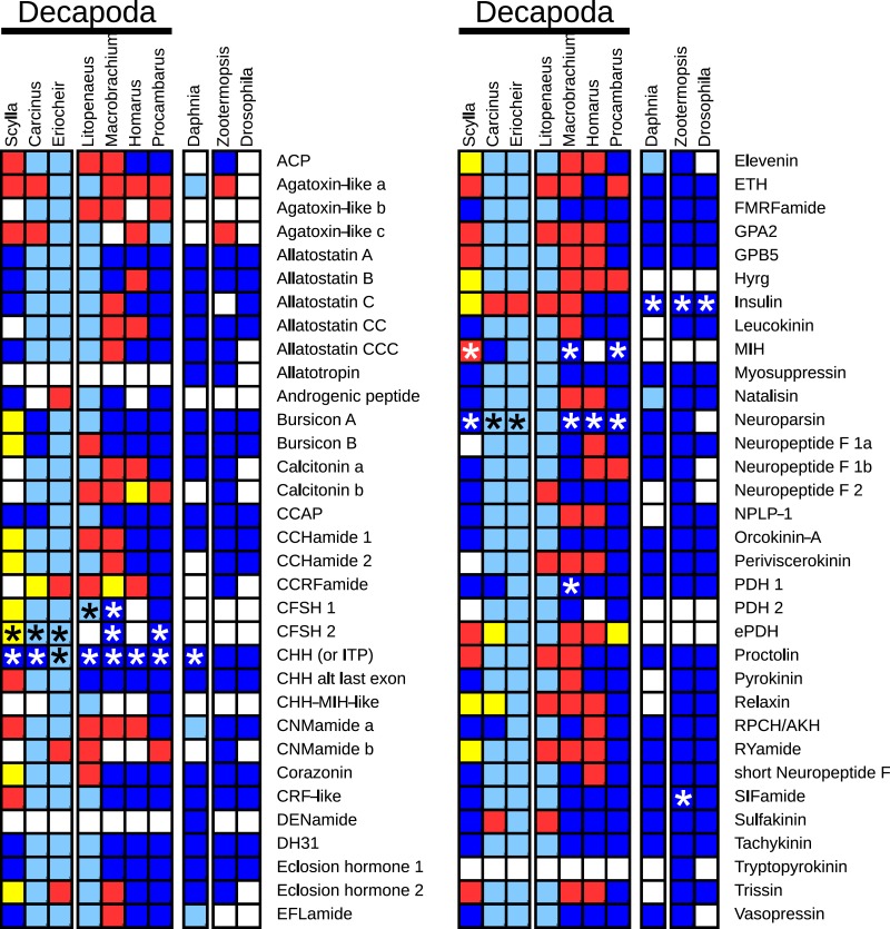
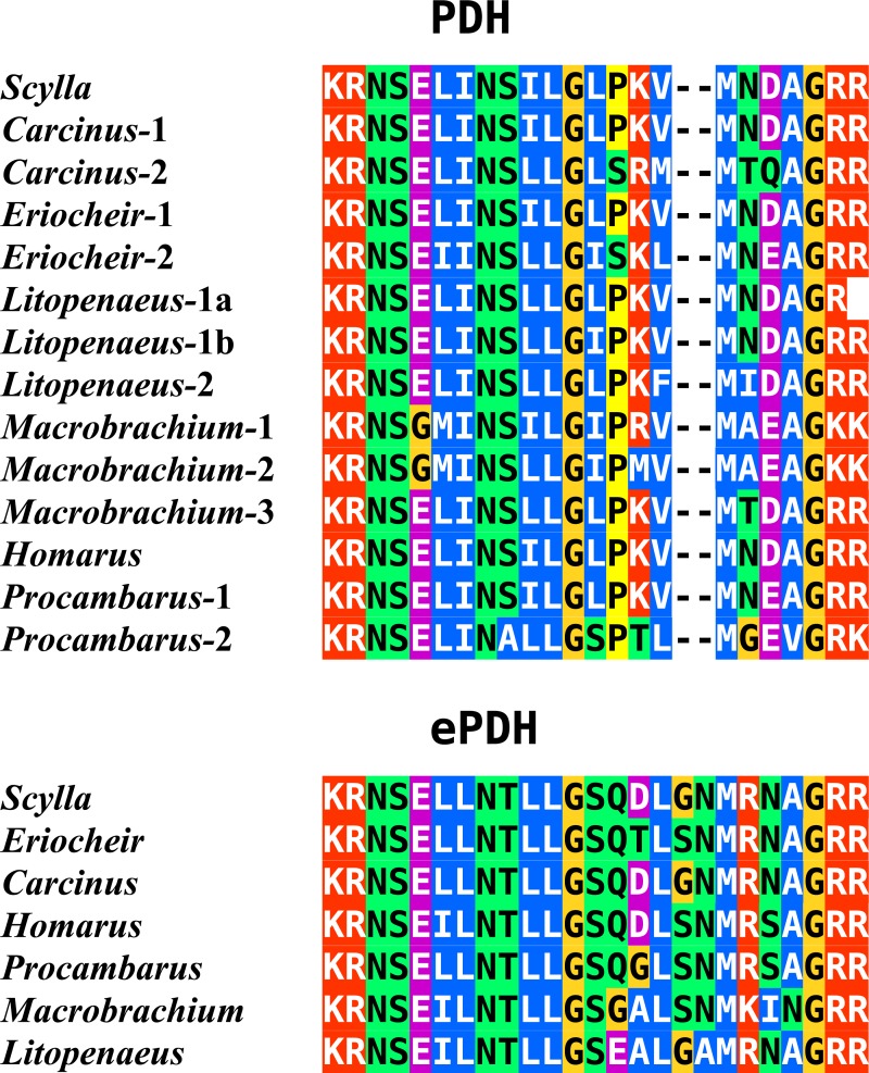
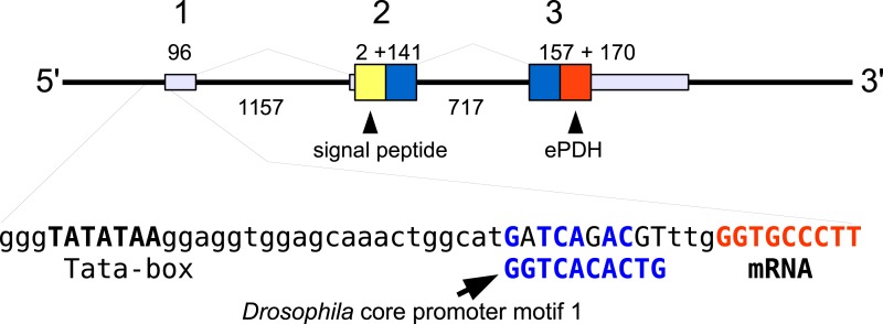
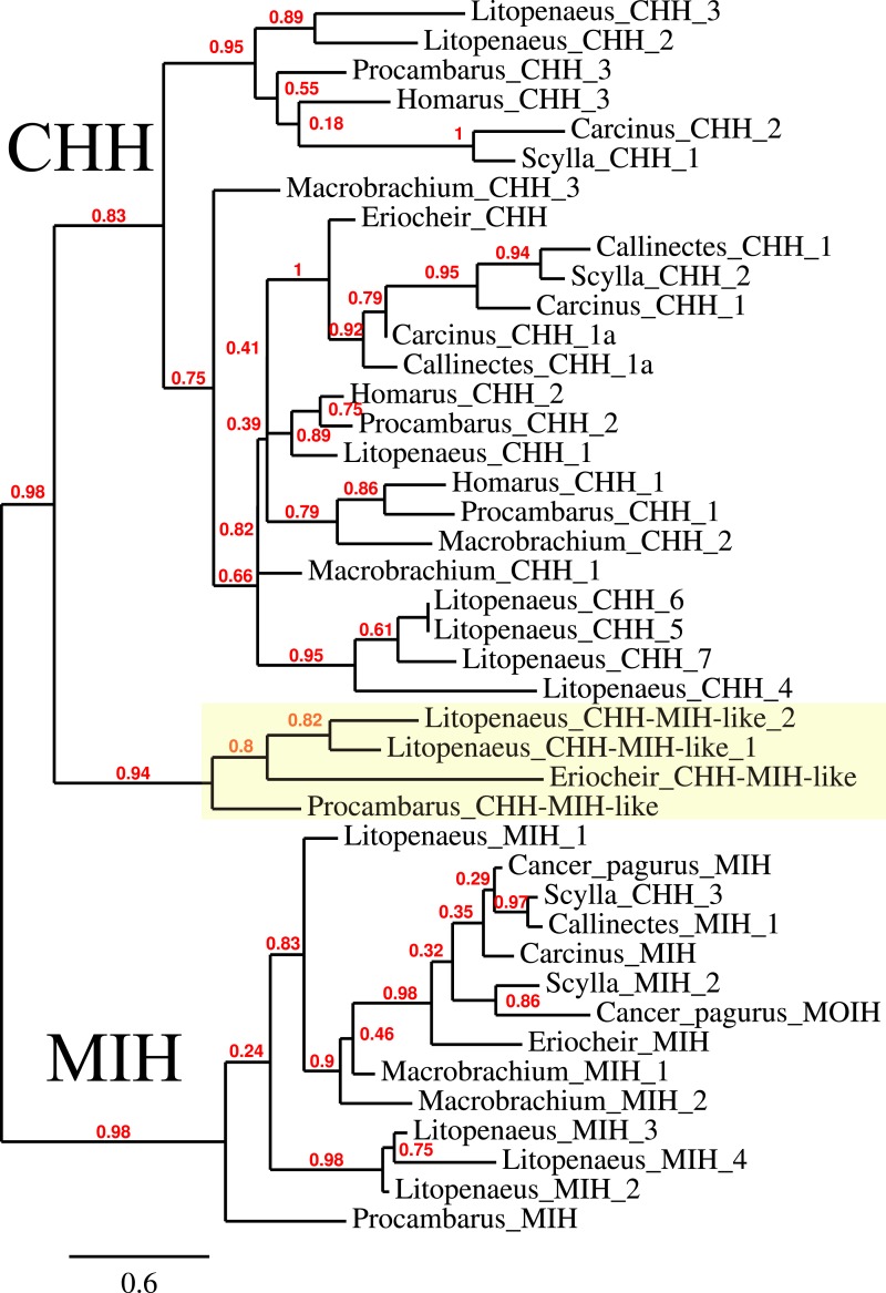
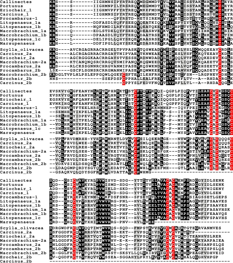
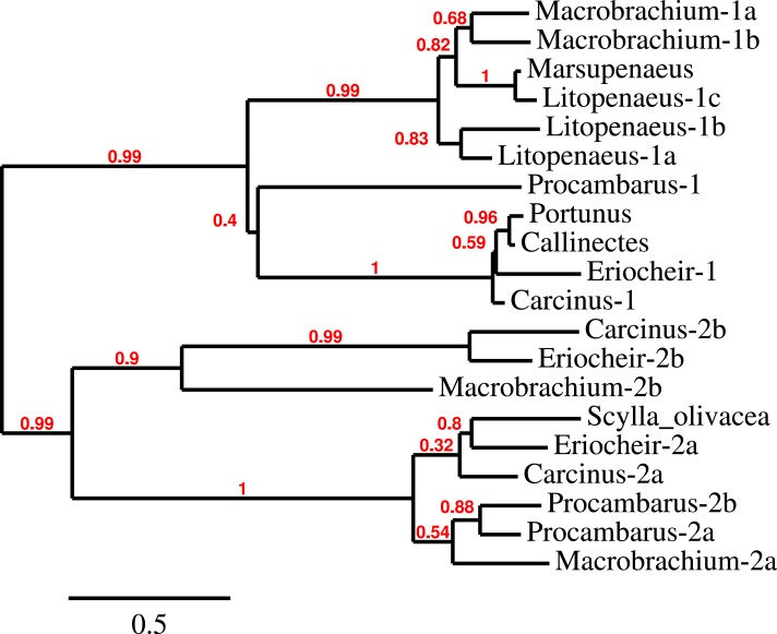
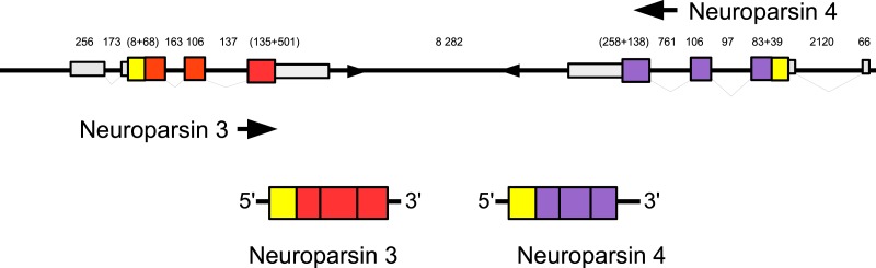
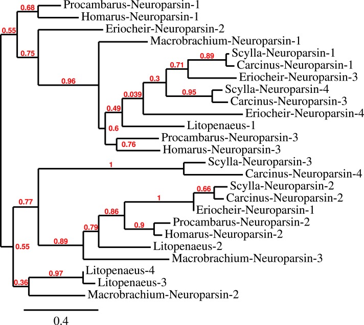
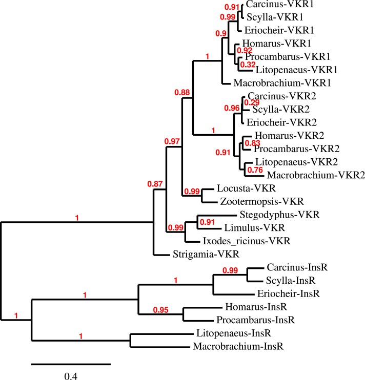
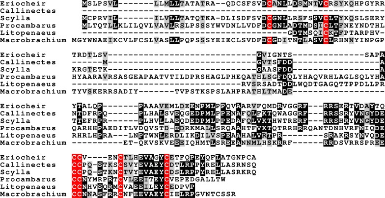
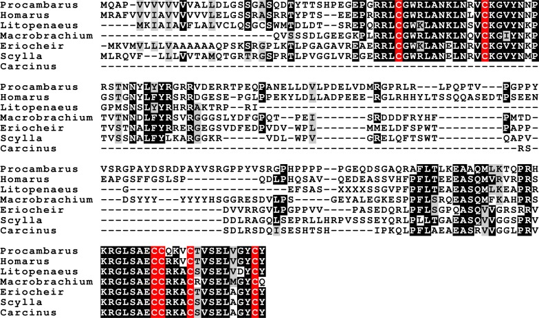
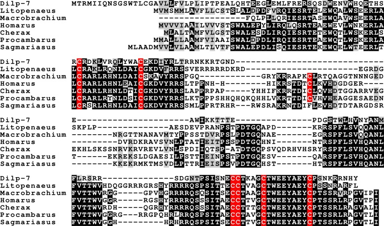
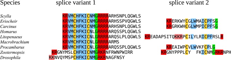
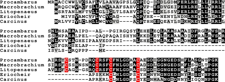
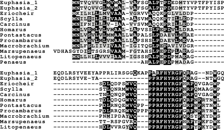
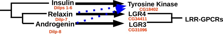
Background. Neuropeptides are important regulators of physiological processes and behavior. Although they tend to be generally well conserved, recent results using trancriptome sequencing on decapod crustaceans give the impression of significant differences between species, raising the question whether such differences are real or artefacts. Methods. The BLAST+ program was used to find short reads coding neuropeptides and neurohormons in publicly available short read archives. Such reads were then used to find similar reads in the same archives, and the DNA assembly program Trinity was employed to construct contigs encoding the neuropeptide precursors as completely as possible. Results. The seven decapod species analyzed in this fashion, the crabs Eriocheir sinensis, Carcinus maenas and Scylla paramamosain, the shrimp Litopenaeus vannamei, the lobster Homarus americanus, the fresh water prawn Macrobrachium rosenbergii and the crayfish Procambarus clarkii had remarkably similar neuropeptidomes. Although some neuropeptide precursors could not be assembled, in many cases individual reads pertaining to the missing precursors show unambiguously that these neuropeptides are present in these species. In other cases, the tissues that express those neuropeptides were not used in the construction of the cDNA libraries. One novel neuropeptide was identified: elongated PDH (pigment dispersing hormone), a variation on PDH that has a two-amino-acid insertion in its core sequence. Hyrg is another peptide that is ubiquitously present in decapods and is likely a novel neuropeptide precursor. Discussion. Many insect species have lost one or more neuropeptide genes, but apart from elongated PDH and hyrg all other decapod neuropeptides are present in at least some insect species, and allatotropin is the only insect neuropeptide missing from decapods. This strong similarity between insect and decapod neuropeptidomes makes it possible to predict the receptors for decapod neuropeptides that have been deorphanized in insects. This includes the androgenic insulin-like peptide that seems to be homologous to drosophila insulin-like peptide 8.

Keywords: Agatoxin-like peptide; Androgenic insulin-like peptide; Calcitonin; Crustacean female sex hormone; Evolution; Neuroparsin; Neuropeptide; PDH; Receptor.


Conflict of interest statement

The author declares there are no competing interests.

Figures

Figure 1
Figure 1. Overview of the presence of neuropeptide genes in seven decapods, Daphnia pulex and two insect species.
Dark blue, neuropeptide precursors that have been published previously; light blue, neuropeptide precursors (or significant parts therefore) that can be deduced directly from publicly available TSAs; red, precursors assembled here; yellow, precursors that could not be assembled, but for which individual reads in TSAs demonstrate their existence in the particular species. Asterisks indicate the existence of more than one gene.
Figure 2
Figure 2. Sequence alignment of PDH and ePDH.
Parts of the various PDH precursors including the convertase cleavage sites of the various decapod species. Note that the ubiquitous presence of ePDH that has a two-amino-acid insertion.
Figure 3
Figure 3. Structure of the ePDH gene from Eriocheir sinensis.
The ePDH gene consists of three exons and two introns. DNA sequences coding the signal peptide in yellow, mature ePDH sequence in red and the remainder of the precursor in blue. Numbers indicate sizes of introns and exons in nucleotides. The DNA sequence containing the TATA box and a sequence that is recognizably similar to the Drosophila core promoter motif 1 (in blue, Ohler, 2006) and the start of the mRNA (in red) are also displayed; the red nucleotides at the end are part of the mRNA.
Figure 4
Figure 4. Phylogenetic tree showing the evolutionary relationships between the CHH and MIH hormones.
Hormones are those identified from decapod SRAs as well as a few for which the biological activity has been described. Highlighted in yellow are the four sequences that on the tree are more similar to CHH, but lack the precursor-related peptide typically present in CHH.
Figure 5
Figure 5. CFSH alignments.
Note that the two types of CFSH are clearly different from one another. Scylla paramamosain is not included in the figure, as there are only a few individual reads from this species. These reads correspond to orthologs of the three crab CFSHs; those that correpond to S. paramamosain 2a predict a protein sequence that is completely identical to the S. olivacea sequence illustrated.
Figure 6
Figure 6. CFSH phylogenetic tree.
Phylogenetic tree of the various CFSH orthologs identified here and elsewhere. The only Scylla sequence is from S. olivacea (GDRN01022056.1). S. paramosain has a very limited number of SRA reads that correspond to three orthologs found in Carcinus and Eriocheir. Note that Macrobrachium, Litopenaeus and Procambarus seem to have independently gone through relatively recent gene duplications.
Figure 7
Figure 7. Configuration of Eriocheir neuroparsin genes 3 and 4.
The relative organization of the two neuroparsin genes relative to one another is indicated. The two genes are located on opposite strands and each gene has four exons and three introns. Numbers indicate the lengths of the exons, introns and the intergenic distance in nucleotides.
Figure 8
Figure 8. Neuroparsin phylogenetic tree.
The different decapod neuroparsin sequences found in the different species were used to make a phylogenetic tree. Note that one neuroparsin gene duplication likely occurred after the crabs separated from the other decapods.
Figure 9
Figure 9. Phylogenetic tree of the tyrosine kinase domains of the decapod insulin and venus kinase receptors.
Venus kinase receptors from the following species were added for increased resolution: Limulus polyphemus, Stegodyphus mimosarum, Locusta migratoria, Ixodes ricinus and Zootermopsis nevadensis. Note that the duplication of the venus kinase receptor gene is not generally present in arthropods and could thus be specific to crustaceans.
Figure 10
Figure 10. Sequence alignment of the decapod adrogenic insulin-like peptides.
Note the relatively poor conservation of the primary sequences of these hormones. Conserved residues indicated in black highlighting, and cysteine residues in red.
Figure 11
Figure 11. Sequence alignment of the decapod insulin-like peptides.
Note the much better conservation of the primary sequences of the A and B chains of these hormones. The Carcinus sequence, although incomplete, is clearly part of an insulin precursor. Conserved residues indicated in black highlighting, and cysteine residues in red.
Figure 12
Figure 12. Sequence alignment of the decapod relaxins.
Note the relatively good sequences conservation between the different decapod peptides and Dilp-7. Conserved residues indicated in black highlighting, and cysteine residues in red.
Figure 13
Figure 13. Last parts of CNMamide precursors.
Some arthropods produce alternatively spliced mRNA predicted to produce different CNMamides. Notice that the major splice variants produce a much better conserved neuropeptide than the alternative ones. Residues in red are predicted to be cleaved by convertase and removed by carboxypeptidase during processing; the green glycine residues will be transformed in C-terminal amides and the cysteine residues are orange. Residues conserved between the different species are in blue.
Figure 14
Figure 14. Sequence alignment of the decapod B-calcitonins.
Some of the decapod B-calcitonins are predicted to have two cysteine bridges in the N-terminal part of their sequence, rather than one.
Figure 15
Figure 15. Hyrg sequence alignment.
Note that only a small part of the sequence of this puative neuropeptide is conserved in both decapods as well as in Euphausia crystallorophias.
Figure 16
Figure 16. Ligand–receptor interactions of insulin-related peptides.
Figure indicates the postulated major interactions of the three decapod insulin-like peptides with three receptors. Secondary interactions are indicated by broken lines. Drosophila gene numbers for orthologous genes are indicated in red. LRR-GPCRs, Leucine-riche repeat GPCRs.

References

    1. Adhama IM, Emmen JMA, Engel W. The role of the testicular factor INSL3 in establishing the gonadal position. Molecular and Cellular Endocrinology. 2000;160:11–16. doi: 10.1016/S0303-7207(99)00188-4. - DOI - PubMed
    1. Aizen J, Chandler JC, Fitzgibbon QP, Sagi A, Battaglene SC, Elizur A, Ventura T. Production of recombinant insulin-like androgenic gland hormones from three decapod species: in vitro testicular phosphorylation and activation of a newly identified tyrosine kinase receptor from the Eastern spiny lobster, Sagmariasus verreauxi. General and Comparative Endocrinology. 2016;229:8–18. doi: 10.1016/j.ygcen.2016.02.013. - DOI - PubMed
    1. Alfa RW, Park S, Skelly KR, Poffenberger G, Jain N, Gu X, Kockel L, Wang J, Liu Y, Powers AC, Kim SK. Suppression of insulin production and secretion by a decretin hormone. Cell Metabolism. 2015;21:323–333. doi: 10.1016/j.cmet.2015.01.006. - DOI - PMC - PubMed
    1. Badisco L, Marchal E, Van Wielendaele P, Verlinden H, Vleugels R, Vanden Broeck J. RNA interference of insulin-related peptide and neuroparsins affects vitellogenesis in the desert locust Schistocerca gregaria. Peptides. 2011;32:573–580. doi: 10.1016/j.peptides.2010.11.008. - DOI - PubMed
    1. Bao C, Yang Y, Huang H, Ye H. Neuropeptides in the cerebral ganglia of the mud crab, Scylla paramamosain: transcriptomic analysis and expression profiles during vitellogenesis. Scientific Reports. 2015;5 doi: 10.1038/srep17055. Article 17055. - DOI - PMC - PubMed

LinkOut - more resources