

DICOM for quantitative imaging biomarker development: a standards based approach to sharing clinical data and structured PET/CT analysis results in head and neck cancer research
- PMID: 27257542
- PMCID: PMC4888317
- DOI: 10.7717/peerj.2057
DICOM for quantitative imaging biomarker development: a standards based approach to sharing clinical data and structured PET/CT analysis results in head and neck cancer research
Abstract
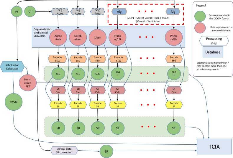
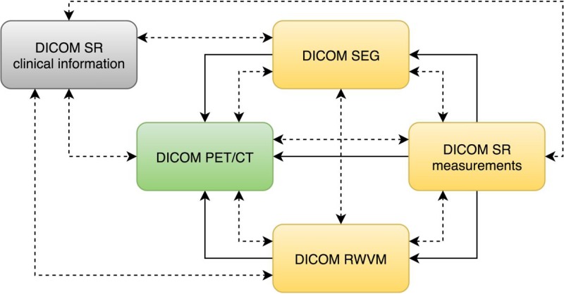
Background. Imaging biomarkers hold tremendous promise for precision medicine clinical applications. Development of such biomarkers relies heavily on image post-processing tools for automated image quantitation. Their deployment in the context of clinical research necessitates interoperability with the clinical systems. Comparison with the established outcomes and evaluation tasks motivate integration of the clinical and imaging data, and the use of standardized approaches to support annotation and sharing of the analysis results and semantics. We developed the methodology and tools to support these tasks in Positron Emission Tomography and Computed Tomography (PET/CT) quantitative imaging (QI) biomarker development applied to head and neck cancer (HNC) treatment response assessment, using the Digital Imaging and Communications in Medicine (DICOM(®)) international standard and free open-source software. Methods. Quantitative analysis of PET/CT imaging data collected on patients undergoing treatment for HNC was conducted. Processing steps included Standardized Uptake Value (SUV) normalization of the images, segmentation of the tumor using manual and semi-automatic approaches, automatic segmentation of the reference regions, and extraction of the volumetric segmentation-based measurements. Suitable components of the DICOM standard were identified to model the various types of data produced by the analysis. A developer toolkit of conversion routines and an Application Programming Interface (API) were contributed and applied to create a standards-based representation of the data. Results. DICOM Real World Value Mapping, Segmentation and Structured Reporting objects were utilized for standards-compliant representation of the PET/CT QI analysis results and relevant clinical data. A number of correction proposals to the standard were developed. The open-source DICOM toolkit (DCMTK) was improved to simplify the task of DICOM encoding by introducing new API abstractions. Conversion and visualization tools utilizing this toolkit were developed. The encoded objects were validated for consistency and interoperability. The resulting dataset was deposited in the QIN-HEADNECK collection of The Cancer Imaging Archive (TCIA). Supporting tools for data analysis and DICOM conversion were made available as free open-source software. Discussion. We presented a detailed investigation of the development and application of the DICOM model, as well as the supporting open-source tools and toolkits, to accommodate representation of the research data in QI biomarker development. We demonstrated that the DICOM standard can be used to represent the types of data relevant in HNC QI biomarker development, and encode their complex relationships. The resulting annotated objects are amenable to data mining applications, and are interoperable with a variety of systems that support the DICOM standard.
Keywords: Cancer imaging; DICOM; Head and neck cancer; Image analysis; Imaging biomarker; Imaging informatics; Interoperability; Open science; PET/CT imaging; Quantitative imaging.
Conflict of interest statement
David Clunie is the owner of PixelMed Publishing, LLC, Bangor, Pennsylvania, USA; Michael Onken is an employee of Open Connections GmbH; Jörg Riesmeier is a freelancer in Oldenburg, Germany; Steve Pieper is an employee of Isomics, Inc., Cambridge, Massachusetts, USA; and Ron Kikinis is an employee of Fraunhofer MEVIS, Bremen, Germany. The contents are solely the responsibility of the authors and do not necessarily represent the official views of the NCI/NIH.
Figures
References
-
- Beichel RR, Van Tol M, Ulrich EJ, Bauer C, Chang T, Plichta KA, Smith BJ, Sunderland JJ, Graham MM, Sonka M, Buatti JM. Semi-automated segmentation of head and neck cancers in 18F-FDG PET scans: a just-enough-interaction approach. Medical Physics. 2016;43(6):2948. doi: 10.1118/1.4948679. - DOI - PMC - PubMed
-
- Bidgood WD., Jr Documenting the information content of images. Proceedings: a conference of the American Medical Informatics Association/AMIA annual fall symposium. AMIA fall symposium; 1997. pp. 424–428. Available at http://www.ncbi.nlm.nih.gov/pmc/articles/PMC2233520/ - PMC - PubMed
-
- Bidgood WD., Jr The SNOMED DICOM microglossary: controlled terminology resource for data interchange in biomedical imaging. Methods of Information in Medicine. 1998;37:404–414. - PubMed
-
- Bidgood WD, Jr, Korman LY, Golichowski AM, Hildebrand PL, Mori AR, Bray B, Brown NJG, Spackman KA, Dove SB, Schoeffler K. Controlled terminology for clinically-relevant indexing and selective retrieval of biomedical images. International Journal on Digital Libraries. 1997;1:278–287. doi: 10.1007/s007990050022. - DOI
Grants and funding
LinkOut - more resources
Full Text Sources
Other Literature Sources
Molecular Biology Databases
