

Effect of fibrin glue on corneal lamellar healing and how it correlates to biomechanical properties: biomechanical wavefront analysis and confocal study
- PMID: 27257609
- PMCID: PMC4890498
- DOI: 10.1186/s40662-016-0046-6
Effect of fibrin glue on corneal lamellar healing and how it correlates to biomechanical properties: biomechanical wavefront analysis and confocal study
Abstract
Background: To evaluate, using a rabbit model, the influence of the wound healing process at the flap edge on corneal biomechanics after sutured, glued, and non-augmented microkeratome flaps.
Methods: Unilateral 160 μm thick laser in situ keratomileusis (LASIK) flaps using a mechanical microkeratome were performed on the corneas of the left eyes of 36 rabbits. Animals were then divided into 3 groups of 12 rabbits each: A: the flaps were glued with human fibrin tissue adhesive (Tisseel); B: the flaps were sutured; and C: the flaps were allowed to heal without the use of sutures or glue (non-augmented). The contralateral eyes served as controls. Reichert ocular response analyzer (ORA) was used to measure corneal hysteresis (CH), corneal resistance factor (CRF), Goldmann-correlated intraocular pressure (IOPg) and cornea-compensated IOP (IOPcc) at 6 weeks and 3 months postoperatively. In vivo confocal microscopy (IVCM) was also used to study the corneal wound healing process in all groups.
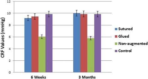
Results: Both mean CH and mean CRF were significantly higher in sutured and glued groups compared with the non-augmented group at 6 weeks and 3 months postoperatively (P < 0.0001). No statistically significant difference in corneal biomechanics was found between controls and groups A and B at any time points. Activated keratocytes were detected at the wound edge and peripheral flap interface in sutured and glued groups.
Conclusion: The healing process at the wound edge is critical for optimal corneal integrity. Fibrin glue may serve as a safe and effective substitute to sutures in enhancing the corneal flap edge healing response and in increasing its mechanical strength.
Keywords: Confocal microscopy; Corneal hysteresis; Corneal resistance factor; Ocular Response Analyzer (ORA); Wound healing.
Figures


References
LinkOut - more resources
Full Text Sources
Other Literature Sources
