. 2016 May 24;3(2):ENEURO.0047-16.2016.
doi: 10.1523/ENEURO.0047-16.2016. eCollection 2016 Mar-Apr.

c-Jun N-Terminal Phosphorylation: Biomarker for Cellular Stress Rather than Cell Death in the Injured Cochlea

Affiliations

c-Jun N-Terminal Phosphorylation: Biomarker for Cellular Stress Rather than Cell Death in the Injured Cochlea

Tommi Anttonen et al. eNeuro. .

Abstract

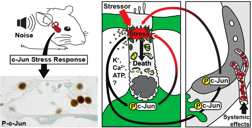
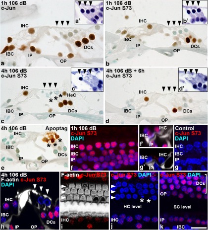
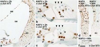
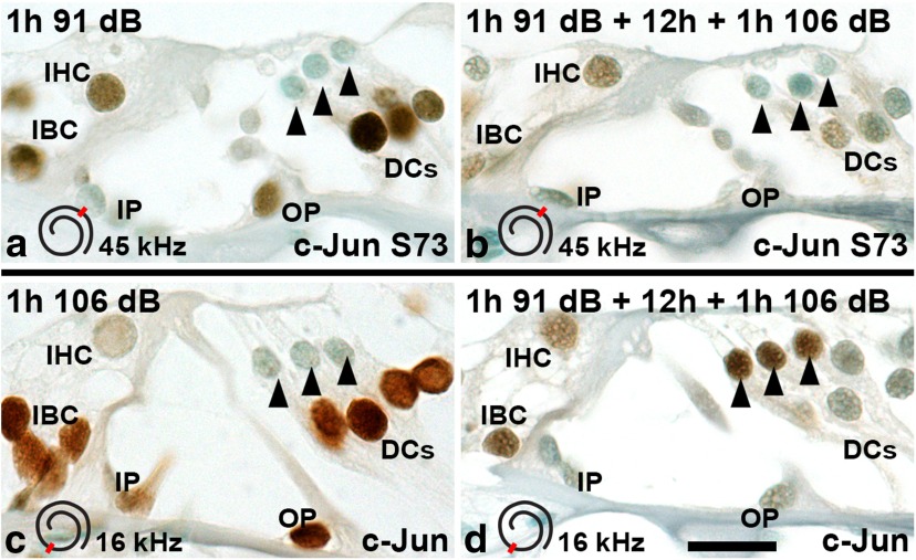
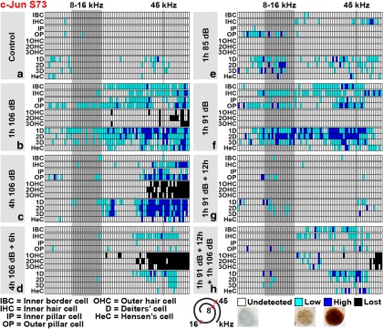
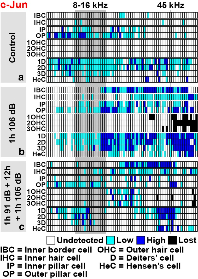
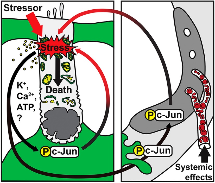
Prevention of auditory hair cell death offers therapeutic potential to rescue hearing. Pharmacological blockade of JNK/c-Jun signaling attenuates injury-induced hair cell loss, but with unsolved mechanisms. We have characterized the c-Jun stress response in the mouse cochlea challenged with acoustic overstimulation and ototoxins, by studying the dynamics of c-Jun N-terminal phosphorylation. It occurred acutely in glial-like supporting cells, inner hair cells, and the cells of the cochlear ion trafficking route, and was rapidly downregulated after exposures. Notably, death-prone outer hair cells lacked c-Jun phosphorylation. As phosphorylation was triggered also by nontraumatic noise levels and none of the cells showing this activation were lost, c-Jun phosphorylation is a biomarker for cochlear stress rather than an indicator of a death-prone fate of hair cells. Preconditioning with a mild noise exposure before a stronger traumatizing noise exposure attenuated the cochlear c-Jun stress response, suggesting that the known protective effect of sound preconditioning on hearing is linked to suppression of c-Jun activation. Finally, mice with mutations in the c-Jun N-terminal phosphoacceptor sites showed partial, but significant, hair cell protection. These data identify the c-Jun stress response as a paracrine mechanism that mediates outer hair cell death.

Keywords: c-Jun phosphorylation; cell death; hair cell; inner ear; noise; supporting cell.


Conflict of interest statement

The authors declare no conflicting financial interests.

Figures

None
Graphical abstract
Figure 1.
Figure 1.
c-Jun expression and S73 phosphorylation during development. a, At P0, tympanic border cells and scattered cells of the lateral wall and Reissner’s membrane express c-Jun. Inset reveals that this expression is not linked with c-Jun S73 phosphorylation, shown in the tympanic border cell region. b, At P6, c-Jun is expressed in the greater epithelial ridge and in Schwann cells of the cochlear nerve (arrow). b', Schematic representation of the organ of Corti at P6: greater epithelial ridge (red), inner hair cell (blue), outer hair cells (cyan), inner pillar cell (yellow), outer pillar cell (orange), Deiters’ cells (green), and Hensen cell (gray). c, c-Jun S73 is phosphorylated in the greater epithelial ridge at P6. Arrowheads mark outer hair cells. c', An adjacent, ApopTag-stained section shows apoptotic fragments in the same area. d, At P0, the annular ligament of stapes shows c-Jun S73 phosphorylation. d', Apoptotic cells are not present in this structure, as evidenced by cleaved caspase-3 staining. e, At P12, c-Jun is expressed in Deiters’ cells of the organ of Corti and in the root cell area of the lateral wall. e', Schematic representation of the cytoarchitecture of the mature organ of Corti: inner border cells (magenta). Other color codes as in b'. AL, Annular ligament of stapes; apop, ApopTag-staining; casp, cleaved caspase-3; DC, Deiters’ cell; IP, inner pillar cell; OC, organ of Corti; OP, outer pillar cell; TBC, tympanic border cell; RC, root cell; RM, Reissner’s membrane; S, stapes; SV, stria vascularis; GER, greater epithelial ridge. Scale bar: (in d') a, a', b, d, d', e, 40 µm; (in d') c, c', 10 µm.
Figure 2.
Figure 2.
The c-Jun stress response in Gfi1-depleted developing hair cells. a, Cochlear hair cells of heterozygous Gfi1GFP/+ mice at P0 have normal morphology, revealed by myosin 7a immunostaining. Arrowheads mark outer hair cells. a', c-Jun S73 immunoreactivity is not detected in the organ of Corti of these control animals. b, In homozygous Gfi1GFP/GFP mice, hair cell morphology is severely distorted. c, d, c-Jun is expressed and phosphorylated in the organ of Corti of these mutants. Note that tympanic border cells show basal c-Jun expression, but its S73 phosphorylation is absent. d', Cleaved caspase-3-positive apoptotic fragments in the hair cell region, shown in an adjacent section. e, In cochleas of Gfi1GFP/+ mice, the GFP reporter marks hair cells. Boxed areas indicate the location of images in f and g. f, In the medial turn of these control specimens, GFP-positive hair cells (arrowheads) lack c-Jun S73 phosphorylation. g, Also in the basal turn, where cellular differentiation is more advanced, GFP-positive hair cells lack the S73 signal. It is also absent from Sox2-positive supporting cells. h, In cochleas of Gfi1GFP/GFP mice, the medial turn shows scattered hair cell loss, while this loss is more severe in the basal turn, consistent with the basal-to-medial-turn progression of the death of Gfi1-depleted hair cells. Boxed areas indicate the location of images in i and j. i, In the medial turn, the morphology of GFP-positive outer hair cells is distorted, and c-Jun is phosphorylated in these degenerating cells. GFP-positive apoptotic fragments (asterisk) can be seen. j, Only a few GFP-positive hair cells are left in the basal turn. Note that the GFP-positive hair cells lacking c-Jun S73 phosphorylation (arrows) are morphologically quite intact. Sox2-expressing supporting cells lack c-Jun S73 immunoreactivity. b, Basal turn; casp, cleaved caspase-3; m, medial turn; TBC, tympanic border cell; IHCs, inner hair cells; GER, greater epithelial ridge. Scale bar: (in d') a–d', 10 µm; (in d') e, h, 385 µm; (in d') f, g, i, j, 15 µm.
Figure 3.
Figure 3.
Acoustic overstimulation triggers an acute and transient c-Jun stress response in the potassium-recycling route of the adult cochlea. a, Scattered pillar and Deiters’ cells of the adult organ of Corti express c-Jun, together with a few cells in the lateral wall. b, c-Jun is only sporadically phosphorylated in cells of the nontraumatized organ of Corti and lateral wall. c, d, Noise exposure for 1 h at the level of 106 dB SPL triggers the upregulation of c-Jun expression and S73 phosphorylation in the organ of Corti and lateral wall. c', d', In the lateral wall, c-Jun activation is most prominent in the root cell area. e, Four hours of noise at 106 dB SPL shifts S73 phosphorylation in the lateral wall to stria vascularis and underlying fibrocytes. e', Closer examination shows that c-Jun is phosphorylated mainly in basal cells, in addition to fibrocytes. Strial cell layers are depicted and color coded above the histological image: Basal cells (blue); intermediate cells (yellow) together with endothelial cells lining capillaries (red); border cells (green). e'', In the basalmost, most damaged part of the cochlea, border cells and intermediate cells of stria vascularis also show S73 phosphorylation. f–g', Following the acute induction, the c-Jun stress response largely disappears from the organ of Corti and lateral wall between 6 and 22 h. h, Schematic presentation of the suggested potassium-recycling route in the cochlea. Color codes: outer hair cells (blue); Deiters’, Hensen, Claudius, and root cells (green); fibrocytes (yellow); stria vascularis (red). i, j, Noise exposure fails to upregulate c-Jun expression and its phosphorylation in nonsensory cells of Gfi1-depleted cochleas that lack most hair cells. All panels except e'' are from the 45–50 kHz region of the cochlea. Fi, Fibrocytes; OC, organ of Corti; RC, root cell; SM, scala media; SV, stria vascularis. Scale bar: (in j) a–g, i, j, 20 µm; (in j) c', d', e', e'', f', g', 45 µm.
Figure 4.
Figure 4.
Acoustic overstimulation triggers the c-Jun stress response in the organ of Corti. a–d, Dynamics of c-Jun expression and S73 phosphorylation in the organ of Corti after noise exposure, which are shown in paraffin sections prepared at various postexposure time points. Arrowheads mark outer hair cells. The c-Jun stress response is restricted to nonsensory cells and inner hair cells, while it is absent in vulnerable outer hair cells. Note the seemingly intact, degenerating or lost status of outer hair cells at various time points. Dying outer hair cells are visible (c, asterisks). Note also that c-Jun phosphorylation is particularly transient in inner border cells around inner hair cells, as these cells are not anymore phosphorylated at the end of the 4-h-long noise exposure. See Results for details. a', b', c', d', Degeneration status of outer hair cells is shown in hematoxylin-stained sections that are consecutive to the sections prepared for immunohistochemistry. e, Outer hair cells show prominent ApopTag staining immediately after noise exposure. f, Consistent with paraffin sections, whole-mount specimens imaged under confocal microscopy show c-Jun S73 induction in inner hair cells, inner border cells, and inner pillar cells acutely after a 1-h-long noise exposure. f', Side view of the corresponding confocal stack. DAPI stains cell nuclei. Phalloidin labels the F-actin cytoskeleton of pillar cells. g, g', In corresponding views from nonexposed specimens, c-Jun S73 expression is absent. h, Consistent with paraffin sections, immediately after a 4-h-long exposure, inner hair cells as well as supporting cell populations, except for inner border cells, are positive for the c-Jun S73 signal, shown in a side view. i, j, Immediately after a 4-h-long exposure, a surface view from the level of hair cells shows that neither surviving outer hair cells nor these cells with apoptotic profiles (asterisks) express phosphorylated c-Jun. Note that nuclear fragmentation is located in the area where the F-actin-rich apical domain of the corresponding outer hair cell is missing. Inner hair cells do show c-Jun S73 staining. Arrowheads mark the three outer hair cell rows. k, Surface view from the supporting cell level of the same specimen shows c-Jun phosphorylation in Deiters’ and pillar cells, but not anymore in inner border cells. DC, Deiters’ cell; HeC, Hensen’s cell; HC, hair cell; IBC, inner border cell; IP, inner pillar cell; OP, outer pillar cell; SC, supporting cell. Scale bar: (in k) a–k, 10 µm.
Figure 5.
Figure 5.
Ototoxic trauma triggers a robust c-Jun stress response in the cochlea. a, Two hours after ototoxin injections, widespread c-Jun S73 immunoreactivity is seen throughout the lining of scala media, in all three layers of stria vascularis, and in fibrocytes and root cells of the lateral wall, in addition to the organ of Corti. b, c, In the organ of Corti, supporting cells and inner hair cells show c-Jun S73 immunoreactivity. Degenerating outer hair cells (b, arrowheads) and their apoptotic remnants (c, asterisks) lack this phosphorylation. d, e, f, In the lateral wall (d, e) and in the organ of Corti (f), the c-Jun stress response is downregulated to low levels by 60 h postexposure. DC, Deiters’ cell; Fi, fibrocyte; IBC, inner border cell; IP, inner pillar cell; KAFU, kanamycin and furosemide; OC, organ of Corti; OP, outer pillar cell; RC, root cells; RM, Reissner’s membrane; SM, scala media; SV, stria vascularis. Scale bar: (in f) a, d–f, 36 µm; b, c, 10 µm.
Figure 6.
Figure 6.
Sound preconditioning attenuates the c-Jun stress response. Immunohistochemical detection of c-Jun expression and S73 phosphorylation in the organ of Corti after various acoustic overexposure paradigms. The spatial location along the cochlear duct of the area displayed is shown in an inset in each image. a, Exposure for 1 h at 91 dB SPL induces c-Jun phosphorylation comparable to exposure to 106 dB (compare with Fig. 4b). b, Sound preconditioning (see Results for details) attenuates c-Jun phosphorylation, most clearly in Deiters’ cells located around the vulnerable outer hair cells. c, In upper levels of the cochlear duct, exposure for 1 h at 106 dB upregulates c-Jun in supporting cells, in a manner similar to that in lower turns of the cochlea (compare with Fig. 4a). d, In upper levels of the cochlear duct, sound preconditioning limits c-Jun upregulation in supporting cells, in a manner similar to that in lower levels (b), but triggers c-Jun expression in intact-appearing outer hair cells (arrowheads). DC, Deiters’ cell; IBC, inner border cell; IP, inner pillar cell; OP, outer pillar cell. Scale bar: (in d) a–d, 10 µm.
Figure 7.
Figure 7.
Cytocochleograms displaying c-Jun S73 phosphorylation in the organ of Corti along the cochlea after various noise paradigms. a–h, For each noise paradigm, a cytocochleogram generated from one cochlea that represents the average expression pattern is shown. The tonotopic organization of the organ of Corti is displayed from left to right (from apical to basal turns). The noise band used (8–16 kHz) is shown as a gray column, similar to the approximate region (45 kHz) from where most histological views of the current study have been obtained. These sites are also marked in the schematic presentation of the spiraling organ of Corti. Abbreviations and color codes are listed below the grids. Representative examples of weak and strong c-Jun S73 immunostaining are shown. See Materials and Methods for details.
Figure 8.
Figure 8.
Cytocochleograms displaying c-Jun expression in the organ of Corti along the cochlea after various noise paradigms. a–c, See Figure legend 7 and Materials and Methods for details. Note that acoustic preconditioning leads to upregulation of c-Jun expression in outer hair cells, but exclusively at the level above the trauma area (c).
Figure 9.
Figure 9.
a–c, Cytocochleograms displaying c-Jun S73 phosphorylation in the organ of Corti along the cochlea after an ototoxic insult. See Figure legend 7 and Materials and Methods for details. a, Two hours after kanamycin and furosemide injections, c-Jun is strongly phosphorylated in supporting cells and inner hair cells. This response is more widespread than the one induced by acoustic trauma (compare with Fig. 7b). b, Intensity of c-Jun S73 immunostaining in supporting cells decreases rapidly, despite the progression of outer hair cell loss toward the apical turn. Inner hair cells still show strong c-Jun S73 signal at 8 h postexposure. c, At 60 h postexposure, outer hair cell loss has reached the apical turn. At this time point, c-Jun S73 is found only in scattered cells in the organ of Corti.
Figure 10.
Figure 10.
JunAA/AA mice are partially protected from noise-induced outer hair cell (OHC) loss. a, b, Histograms show the mean ± SEM of outer hair cell loss in Junwt/wt (dark gray) and JunAA/AA (light gray) mice 16 days after a 6-h-long noise exposure at 110 or 115 dB SPL. At both noise levels, JunAA/AA mice show a statistically significant attenuation of OHC loss (110 dB, *p = 0.01705; 115 dB, *p = 0.02527). The numbers of cochleas used for this analysis are shown. a', b', OHC loss is shown for each individual cochlea used for the histograms in a and b. c–d', Noise-exposed cochleas from Junwt/wt (c, c') and JunAA/AA (d, d') mice embedded in resin and viewed under DIC optics at the 45 kHz region. Arrowheads point OHCs. The same hair cells/sites of lost hair cells are viewed at the surface and nuclear levels. In these views, 3 and 12 OHCs, respectively, are lost in JunAA/AA and Junwt/wt cochleas. Note that epithelial wound healing (scar formation) is comparable in these genotypes. Arrow shows an example of normal scar formation in JunAA/AA mice. e, Paraffin-embedded and hematoxylin-stained cross section from the 45 kHz region of a noise-exposed JunAA/AA cochlea shows unaltered overall morphology. Arrowheads point to OHCs. f, Noise-exposed JunAA/AA cochleas show insignificant c-Jun S73 immunostaining, indicating the specificity of the antibody used. Very weak staining is seen in Deiters’ and pillar cells, where c-Jun is constitutively expressed and is strongly upregulated upon traumas. Scale bar: (in d') a–d', 10 µm; e, f, 65 µm.
Figure 11.
Figure 11.
Hypothetical model of the c-Jun stress response in the traumatized, adult cochlea. The model is based on the pattern of c-Jun N-terminal phosphorylation upon noise and ototoxic exposures. These stressors induce direct stress on outer hair cells (OHCs). Supporting cells (green) are able to perceive this stress, perhaps through factors released by stressed OHCs (Housley and Gale, 2010), leading to c-Jun phosphorylation in supporting cells. c-Jun is sequentially phosphorylated in the lateral wall (right picture). This temporal sequence follows the suggested sequence of potassium ion recycling within the nonsensory cell compartments and fluids of the cochlea. c-Jun phosphorylation in nonsensory cells might induce additional stress (black-to-red arrows) on OHCs through unknown paracrine mechanisms. Once intolerable levels of cellular stress are reached, OHC death is stimulated. By inhibiting c-Jun phosphorylation (JunAA/AA mice), formation of this additional stress on OHCs can be decreased, leading to increased OHC survival. Besides local cochlear events, systemic factors, such as stress hormones, may contribute to the induction of the c-Jun stress response in the cochlea.

References

    1. Anttonen T, Kirjavainen A, Belevich I, Laos M, Richardson WD, Jokitalo E, Brakebusch C, Pirvola U (2012) Cdc42-dependent structural development of auditory supporting cells is required for wound healing at adulthood. Sci Rep 2:978. 10.1038/srep00978 - DOI - PMC - PubMed
    1. Anttonen T, Belevich I, Kirjavainen A, Laos M, Brakebusch C, Jokitalo E, Pirvola U (2014) How to bury the dead: elimination of apoptotic hair cells from the hearing organ of the mouse. J Assoc Res Otolaryngol 15:975-992. 10.1007/s10162-014-0480-x - DOI - PMC - PubMed
    1. Behrens A, Sibilia M, Wagner EF (1999) Amino-terminal phosphorylation of c-Jun regulates stress-induced apoptosis and cellular proliferation. Nat Genet 21:326-329. http://www.nature.com/ng/journal/v21/n3/full/ng0399_326.html - PubMed
    1. Besirli CG, Wagner EF, Johnson EM (2005) The limited role of NH2-terminal c-Jun phosphorylation in neuronal apoptosis: identification of the nuclear pore complex as a potential target of the JNK pathway. J Cell Biol 170:401-411. 10.1083/jcb.200501138 - DOI - PMC - PubMed
    1. Björkblom B, Vainio JC, Hongisto V, Herdegen T, Courtney MJ, Coffey ET (2008) All JNKs can kill, but nuclear localization is critical for neuronal death. J Biol Chem 283:19704-19713. 10.1074/jbc.M707744200 - DOI - PubMed

Publication types

MeSH terms