. 2016 Nov;281(2):617-624.
doi: 10.1148/radiol.2016152508. Epub 2016 Jun 3.

Effects of Microwave Ablation on Arterial and Venous Vasculature after Treatment of Hepatocellular Carcinoma

Affiliations

Effects of Microwave Ablation on Arterial and Venous Vasculature after Treatment of Hepatocellular Carcinoma

Jason Chiang et al. Radiology. 2016 Nov.

Abstract

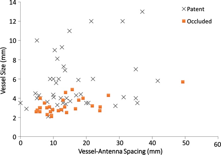
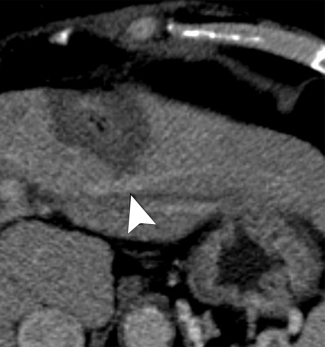
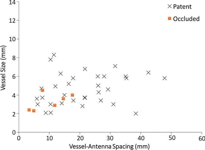
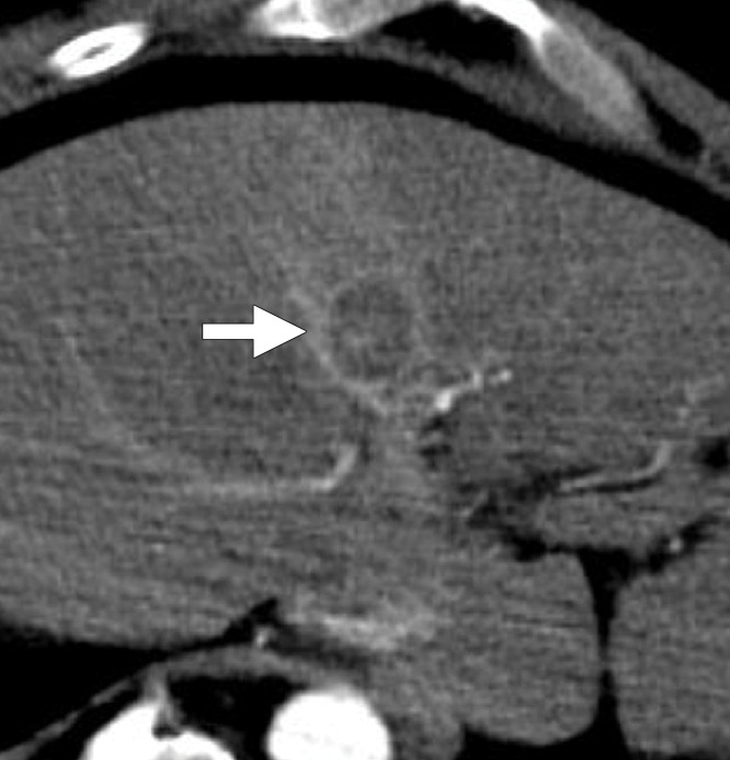
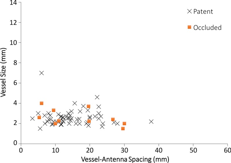
Purpose To characterize vessel occlusion rates and their role in local tumor progression in patients with hepatocellular carcinoma (HCC) who underwent microwave tumor ablation. Materials and Methods This institutional review board approved, HIPAA-compliant retrospective review included 95 patients (75 men and 20 women) with 124 primary HCCs who were treated at a single center between January 2011 and March 2014. Complete occlusion of the portal veins, hepatic veins, and hepatic arteries within and directly abutting the ablation zone was identified with postprocedure contrast material-enhanced computed tomography. For each vessel identified in the ablation zone, its size and antenna spacing were recorded and correlated with vascular occlusion with logistic regression analysis. Local tumor progression rates were then compared between patent and occluded vessels for each vessel type with Fisher exact test. Results Occlusion was identified in 39.7% of portal veins (29 of 73), 15.0% of hepatic veins (six of 40), and 14.2% of hepatic arteries (10 of 70) encompassed within the ablation zone. Hepatic vein occlusion was significantly correlated with a smaller vessel size (P = .036) and vessel-antenna spacing (P = .006). Portal vein occlusion was only significantly correlated with a smaller vessel size (P = .001), particularly in vessels that were less than 3 mm in diameter. Local tumor progression rates were significantly correlated with patent hepatic arteries within the ablation zone (P = .02) but not with patent hepatic (P = .57) or portal (P = .14) veins. Conclusion During microwave ablation of HCC, hepatic veins and arteries were resistant to vessel occlusion compared with portal veins, and only arterial patency within an ablation zone was related to local tumor progression. © RSNA, 2016.


Figures

Figure 1:
Figure 1:
CT image shows measurements of a patent hepatic artery encompassed within a microwave ablation zone (inset). Vessel-antenna spacing (dashed arrow) was measured from the center of the ablation zone (*) to the nearest vessel edge, and vessel size was measured at its widest diameter (arrow) within the ablation zone.
Figure 2a:
Figure 2a:
(a) Axial contrast-enhanced CT image shows an arterially enhancing 2.1-cm HCC in the right hepatic lobe (arrowhead). (b) Axial contrast-enhanced CT image obtained before ablation shows a patent posterior branch of the right portal vein (arrow) approximately 5 mm from the tumor margin. (c) Axial contrast-enhanced CT image obtained immediately after ablation shows portal vein occlusion (arrow) with an associated transient hepatic attenuation difference. (d) Graph shows patent versus occluded portal vein events as a function of vessel size and vessel-antenna spacing. Portal veins that were smaller in size were found to be significantly correlated with occlusion events (P < .001).
Figure 2b:
Figure 2b:
(a) Axial contrast-enhanced CT image shows an arterially enhancing 2.1-cm HCC in the right hepatic lobe (arrowhead). (b) Axial contrast-enhanced CT image obtained before ablation shows a patent posterior branch of the right portal vein (arrow) approximately 5 mm from the tumor margin. (c) Axial contrast-enhanced CT image obtained immediately after ablation shows portal vein occlusion (arrow) with an associated transient hepatic attenuation difference. (d) Graph shows patent versus occluded portal vein events as a function of vessel size and vessel-antenna spacing. Portal veins that were smaller in size were found to be significantly correlated with occlusion events (P < .001).
Figure 2c:
Figure 2c:
(a) Axial contrast-enhanced CT image shows an arterially enhancing 2.1-cm HCC in the right hepatic lobe (arrowhead). (b) Axial contrast-enhanced CT image obtained before ablation shows a patent posterior branch of the right portal vein (arrow) approximately 5 mm from the tumor margin. (c) Axial contrast-enhanced CT image obtained immediately after ablation shows portal vein occlusion (arrow) with an associated transient hepatic attenuation difference. (d) Graph shows patent versus occluded portal vein events as a function of vessel size and vessel-antenna spacing. Portal veins that were smaller in size were found to be significantly correlated with occlusion events (P < .001).
Figure 2d:
Figure 2d:
(a) Axial contrast-enhanced CT image shows an arterially enhancing 2.1-cm HCC in the right hepatic lobe (arrowhead). (b) Axial contrast-enhanced CT image obtained before ablation shows a patent posterior branch of the right portal vein (arrow) approximately 5 mm from the tumor margin. (c) Axial contrast-enhanced CT image obtained immediately after ablation shows portal vein occlusion (arrow) with an associated transient hepatic attenuation difference. (d) Graph shows patent versus occluded portal vein events as a function of vessel size and vessel-antenna spacing. Portal veins that were smaller in size were found to be significantly correlated with occlusion events (P < .001).
Figure 3a:
Figure 3a:
Cirrhosis and HCC in a 61-year-old man. (a) Axial contrast-enhanced CT image obtained after ablation shows a patent left hepatic vein branch (arrowhead) approximately 2.2 cm from the center of the ablation zone. (b) Graph shows patent versus occluded hepatic veins as a function of vessel size and vessel-antenna spacing. Hepatic veins that were smaller in size and had smaller vessel-antenna spacing were found to be significantly correlated with occlusion in terms of both vessel size (P = .036) and vessel-antenna spacing (P = .006).
Figure 3b:
Figure 3b:
Cirrhosis and HCC in a 61-year-old man. (a) Axial contrast-enhanced CT image obtained after ablation shows a patent left hepatic vein branch (arrowhead) approximately 2.2 cm from the center of the ablation zone. (b) Graph shows patent versus occluded hepatic veins as a function of vessel size and vessel-antenna spacing. Hepatic veins that were smaller in size and had smaller vessel-antenna spacing were found to be significantly correlated with occlusion in terms of both vessel size (P = .036) and vessel-antenna spacing (P = .006).
Figure 4a:
Figure 4a:
Hepatitis C infection and HCC in a 64-year-old man. (a) Axial contrast-enhanced CT image obtained immediately after ablation shows that the hepatic artery (arrow) remains patent despite its close proximity (6 mm) to the center of the ablation zone. (b) Graph shows vessel occlusion and patency as a function of hepatic artery size and vessel-antenna spacing. The hepatic arteries thrombosed at a rate similar to that of the hepatic veins (15% vs 14.2%, respectively).
Figure 4b:
Figure 4b:
Hepatitis C infection and HCC in a 64-year-old man. (a) Axial contrast-enhanced CT image obtained immediately after ablation shows that the hepatic artery (arrow) remains patent despite its close proximity (6 mm) to the center of the ablation zone. (b) Graph shows vessel occlusion and patency as a function of hepatic artery size and vessel-antenna spacing. The hepatic arteries thrombosed at a rate similar to that of the hepatic veins (15% vs 14.2%, respectively).

References

    1. Bruix J, Sherman M; American Association for the Study of Liver Diseases . Management of hepatocellular carcinoma: an update. Hepatology 2011;53(3):1020–1022. - PMC - PubMed
    1. European Association For The Study Of The Liver; European Organisation For Research And Treatment Of Cancer . EASL-EORTC clinical practice guidelines: management of hepatocellular carcinoma. J Hepatol 2012;56(4):908–943. - PubMed
    1. Omata M, Lesmana LA, Tateishi R, et al. . Asian Pacific Association for the Study of the Liver consensus recommendations on hepatocellular carcinoma. Hepatol Int 2010;4(2):439–474. - PMC - PubMed
    1. Ahmed M, Brace CL, Lee FT, Jr, Goldberg SN. Principles of and advances in percutaneous ablation. Radiology 2011;258(2):351–369. - PMC - PubMed
    1. Bhardwaj N, Strickland AD, Ahmad F, Atanesyan L, West K, Lloyd DM. A comparative histological evaluation of the ablations produced by microwave, cryotherapy and radiofrequency in the liver. Pathology 2009;41(2):168–172. - PubMed

MeSH terms

LinkOut - more resources