. 2016 Jun 1;5(2):19.
doi: 10.3390/antibiotics5020019.

Insights into the Stress Response Triggered by Kasugamycin in Escherichia coli

Affiliations

Insights into the Stress Response Triggered by Kasugamycin in Escherichia coli

Christian Müller et al. Antibiotics (Basel). .

Abstract

The bacteriostatic aminoglycoside antibiotic kasugamycin inhibits protein synthesis at an initial step without affecting translation elongation. It binds to the mRNA track of the ribosome and prevents formation of the translation initiation complex on canonical mRNAs. In contrast, translation of leaderless mRNAs continues in the presence of the drug in vivo. Previously, we have shown that kasugamycin treatment in E. coli stimulates the formation of protein-depleted ribosomes that are selective for leaderless mRNAs. Here, we provide evidence that prolonged kasugamycin treatment leads to selective synthesis of specific proteins. Our studies indicate that leaderless and short-leadered mRNAs are generated by different molecular mechanisms including alternative transcription and RNA processing. Moreover, we provide evidence for ribosome heterogeneity in response to kasugamycin treatment by alteration of the modification status of the stalk proteins bL7/L12.

Keywords: Escherichia coli; kasugamycin; leaderless mRNA; translation initiation.


Figures

Figure 1
Figure 1
Prolonged Ksg treatment triggers selective protein synthesis. (a) Growth of strain MG1655 harboring plasmid pRB381-1, encoding the leaderless cI-lacZ fusion gene in the absence (black) and presence of 0.5 mg/ml Ksg (red line) was monitored by measuring the optical density at 600 nm. Addition of Ksg is indicated by red arrows. Error bars representing the standard deviation of triplicate samples are hidden by the symbols. (b) At indicated time points before and after addition of Ksg pulse labeling was performed and the labelled proteins were separated by SDS-PAGE. The position of the CI-LacZ fusion protein is indicated by an arrow. (c) Growth of strain MG1655 (solid line) in the absence (black) and presence of 0.5 mg/ml Ksg (red line) was compared to growth of strain MG1655ΔmazF (dotted line) both harboring plasmid pRB381-1. Addition of Ksg is indicated by a red arrow. Again, error bars representing the standard deviation of triplicate samples are hidden by the symbols. (d) At time points indicated pulse labeling was performed and the labelled proteins were separated by SDS-PAGE. The arrow indicates the CI-LacZ fusion protein.
Figure 2
Figure 2
2D gel analysis performed to identify selectively synthesized proteins in the presence of Ksg. (a) and (c) Sections of the 2D gel performed with samples withdrawn from the untreated strain; (b) and (d) the same sections of the 2D gel used for the separation of the proteins labeled in the presence of Ksg. The molecular weight is given to the left and the pH values are indicated at the bottom. The proteins identified are numbered according to Table 1. The position of elongation factor 2 is indicated by an arrow head in (a) and (b) and the position of the unmodified protein uL12 is circled in (d).
Figure 3
Figure 3
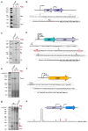
Determination of the 5′-terminus of the rplL (a and b), clpP (c and d), cspA (e and f), and eno (g and h) mRNAs by primer extension analysis. Total RNA was prepared from strain MG1655 before and 120 min after addition of Ksg. Likewise, total RNA was prepared from strain MG1655ΔmazF after treatment with Ksg for 120 min and used for the analysis. The analyses were performed in triplicate and one representative autoradiograph is shown. (b, d, f, and g) Schematic depictions of promoter positions and sequence of 5′-UTRs and proximal coding regions of the different RNAs are shown. Signals corresponding to transcriptional start sites and processing sites are indicted to the right of the primer extension analysis and in the depiction of the sequence. The lengths of the standard marker are given to the left.
Figure 4
Figure 4
Selective translation of the short-leadered ompAΔ117-lacZ mRNA in the presence of Ksg in vivo. (a) Pulse labeling of strain MG1655 harboring plasmid pIM17 grown in the absence or in the presence of Ksg. The arrow indicates the position of the fusion protein. (b) The sequence of the 5′-UTR and the proximal coding region of the mRNA is given. The AUG start codon is underlined and the SD sequence is given in bold.
Figure 5
Figure 5
Formation of RNA43 upon Ksg treatment. Total RNA purified from strain MG1655 (lanes 1–8) and MG1655ΔmazF (lanes 9–16) at indicated time points without (lanes 1–4 and 9–12) or with the addition of Ksg (lanes 5–8 and 13–16) was subjected to northern blot analysis using probe V7, specific for the RNA43. A probe specific for the 5S rRNA was used as an internal control. The second signal observed after addition of Ksg is marked by an asterisk. The obtained RNA43 signal intensities were quantified and normalized using the 5S rRNA signals. In the graph below, the corresponding relative amounts of RNA43 are given Light blue and red bars indicate without or with Ksg treatment, respectively. The value obtained in strain MG1655 at time point 0’ (lane 1) was set to 1. The error bars represent the standard deviation of triplicate samples.

Similar articles

Cited by

References

    1. Okuyama A., Machiyama N., Kinoshita T., Tanaka N. Inhibition by kasugamycin of initiation complex formation on 30s ribosomes. Biochem. Biophys. Res. Commun. 1971;43:196–199. doi: 10.1016/S0006-291X(71)80106-7. - DOI - PubMed
    1. Kozak M., Nathans D. Differential inhibition of coliphage MS2 protein synthesis by ribosome-directed antibiotics. J. Mol. Biol. 1972;70:41–55. doi: 10.1016/0022-2836(72)90162-3. - DOI - PubMed
    1. Poldermans B., Goosen N., Van Knippenberg P.H. Studies on the function of two adjacent N6,N6-dimethyladenosines near the 3′ end of 16 S ribosomal RNA of Escherichia coli. I. The effect of kasugamycin on initiation of protein synthesis. J. Biol. Chem. 1979;254:9085–9089. - PubMed
    1. Woodcock J., Moazed D., Cannon M., Davies J., Noller H.F. Interaction of antibiotics with A- and P-site-specific bases in 16S ribosomal RNA. EMBO J. 1991;10:3099–3103. - PMC - PubMed
    1. Schluenzen F., Takemoto C., Wilson D.N., Kaminishi T., Harms J.M., Hanawa-Suetsugu K., Szaflarski W., Kawazoe M., Shirouzu M., Nierhaus K.H., et al. The antibiotic kasugamycin mimics mRNA nucleotides to destabilize tRNA binding and inhibit canonical translation initiation. Nat. Struct. Mol. Biol. 2006;13:871–878. doi: 10.1038/nsmb1145. - DOI - PubMed

LinkOut - more resources