. 2016 Aug;23(8):1394-405.
doi: 10.1038/cdd.2016.28. Epub 2016 Jun 3.

The long noncoding RNA NRF regulates programmed necrosis and myocardial injury during ischemia and reperfusion by targeting miR-873

Affiliations

The long noncoding RNA NRF regulates programmed necrosis and myocardial injury during ischemia and reperfusion by targeting miR-873

K Wang et al. Cell Death Differ. 2016 Aug.

Abstract

Emerging evidences suggest that necrosis is programmed and is one of the main forms of cell death in the pathological process in cardiac diseases. Long noncoding RNAs (lncRNAs) are emerging as new players in gene regulation. However, it is not yet clear whether lncRNAs can regulate necrosis in cardiomyocytes. Here, we report that a long noncoding RNA, named necrosis-related factor (NRF), regulates cardiomyocytes necrosis by targeting miR-873 and RIPK1 (receptor-interacting serine/threonine-protein kinase 1)/RIPK3 (receptor-interacting serine/threonine-protein kinase 3). Our results show that RIPK1 and RIPK3 participate in H2O2-induced cardiomyocytes necrosis. miR-873 suppresses the translation of RIPK1/RIPK3 and inhibits RIPK1/RIPK3-mediated necrotic cell death in cardiomyocytes. miR-873 reduces myocardial infarct size upon ischemia/reperfusion (I/R) injury in the animal model. In exploring the molecular mechanism by which miR-873 expression is regulated, we identify NRF as an endogenous sponge RNA and repress miR-873 expression. NRF directly binds to miR-873 and regulates RIPK1/RIPK3 expression and necrosis. Knockdown of NRF antagonizes necrosis in cardiomyocytes and reduces necrosis and myocardial infarction upon I/R injury. Further, we identify that p53 transcriptionally activates NRF expression. P53 regulates cardiomyocytes necrosis and myocardial I/R injury through NRF and miR-873.Our results identify a novel mechanism involving NRF and miR-873 in regulating programmed necrosis in the heart and suggest a potential therapeutic avenue for cardiovascular diseases.


Figures
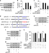
Figure 1
Figure 1
miR-873 participates in the regulation of RIPK1/RIPK3 expression. (a) RIPK1 and RIPK3 expression levels are upregulated upon H2O2 treatment. Cardiomyocytes were treated with 600 μM H2O2 at indicated time. RIPK1 and RIPK3 levels were analyzed by immunoblot (n=3). (b) Knockdown of RIPK1 attenuates necrotic responses induced by H2O2. Cardiomyocytes were infected with adenoviral RIPK1-siRNA or its scramble form (RIPK1-sc), and then treated with 600 μM H2O2. Necrotic cell death was assessed by PI exclusion assay (n=3). *P<0.05 versus H2O2 alone. (c) Knockdown of RIPK3 reduces necrotic responses induced by H2O2. Cardiomyocytes were infected with adenoviral RIPK3-siRNA or its scramble form (RIPK3-sc), and then treated with 600 μM H2O2. Necrotic cell death was assessed by PI assay (n=3). *P<0.05 versus H2O2 alone. (d) Putative miR-873-binding sites in the 3′UTR region of RIPK1 or RIPK3 analyzed by TargetScan program. Mutated miR-873 (miR-873-mut) is shown. (e) miR-873 suppresses the expression of RIPK1 and RIPK3 in cardiomyocytes. Cardiomyocytes were transfected with miR-873 or negative control (NC). RIPK1 and RIPK3 expression levels were analyzed by immunoblot (n=3). (f) Knockdown of miR-873 induces the increase of RIPK1 and RIPK3 expression. Cardiomyocytes were transfected with antagomir miR-873 (anta-873) or its negative control (anta-NC). Forty-eight hours after transfection RIPK1 and RIPK3 expression levels were analyzed by immunoblot (n=3). (g) miR-873 reduces the RIPK1 and RIPK3 expression levels upon H2O2 treatment. Cardiomyocytes were transfected with miR-873 or NC. Twenty-four hours after transfection cells were treated with H2O2. The expression levels of RIPK1 and RIPK3 were analyzed by immunoblot (n=4). (h) Luciferase assay. HEK293 cells were transfected with miR-873 or its mutated form (miR-873-mut), then transfected with the luciferase constructs of the RIPK1 3′UTR or RIPK3 3′UTR. Cells transfected with pGL3 served as a control. The luciferase activity was analyzed (n=3). *P<0.05
Figure 2
Figure 2
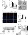
miR-873 regulates H2O2-induced necrosis through RIPK1/RIPK3. (a) The levels of miR-873 in human ischemic myocardium. The control non-ischemic human hearts (normal) and ischemic zones of myocardial infarction hearts (infarction) were used for qRT-PCR analysis of miR-873 levels (n=6). *P<0.05 versus normal. (b) The expression levels of miR-873 were analyzed by qRT-PCR in cardiomyocytes exposed to 600 μM H2O2 at the indicated time (n=4). *P<0.05 versus control. (c) Electron microscopy (EM). Cardiomyocytes were transfected with miR-873 or its negative control (NC). Twenty-four hours after transfection cells were treated with 600 μM H2O2. Representative images showed the characteristics of necrotic cells (n=3). (d) miR-873 reduces necrotic responses induced by H2O2. Cardiomyocytes were treated as described in (c). Necrotic cell death was assessed by PI assay. Representative images showed PI-positive cells (left). Red, PI-positive nuclei; blue, DAPI-stained nuclei. Bar=20 μm. The quantitative analysis of necrosis was shown in the right panel (n=4). *P<0.05 versus H2O2 alone. (e and f) RIPK1/RIPK3 target protectors reduce the inhibitory effect of miR-873 on RIPK1/RIPK3 expression and necrosis. Cardiomyocytes were transfected with miR-873, the target protector (RIPK1-TPmiR-873), the target protector (RIPK3-TPmiR-873) or the control (TPcontrol). (e) RIPK1 and RIPK3 expression were analyzed by immunoblot (n=3). (f) Necrotic cell death was assessed by PI assay (n=3). *P<0.05
Figure 3
Figure 3
miR-873 regulates necrotic cell death in the heart. (a) miR-873 levels during myocardial ischemia/reperfusion (I/R). Mice were subjected to cardiac I/R at the indicated time. The Sham-operated group served as a negative control. Ischemic zone and Remote zone were prepared at the indicated time for qRT-PCR analysis of miR-873 levels (n=6). *P<0.05 versus 0 min or sham. (b) Enforced expression of miR-873 attenuates the increase of RIPK1/RIPK3 levels in response to I/R jury. Intracoronary delivery of miR-873 and I/R is described in Materials and Methods. RIPK1/RIPK3 levels were analyzed by immunoblot (n=4). (c) miR-873 attenuates myocardial necrosis upon I/R injury. In vivo delivery of miR-873 or NC was performed as described in Materials and Methods. The mice were subjected to sham or I/R as described in the Materials and Methods. Myosin antibody was injected into the mice to label necrotic cells. Representative images of ventricular myocardium sections from sham operation or I/R are shown in the left panel and the quantitative analysis of myosin-positive cells is shown in the right panel. Green, immunohistochemistry for myosin antibody incorporation into the heart; red, wheat germ stain for cell membranes; blue, DAPI-stained nuclei (n=6). Bar=20 μm. *P<0.05 versus WT+I/R. (d) Enforced expression of miR-873 attenuates myocardial infarction sizes. Mice were treated as described in (c). Myocardial infarct sizes were measured as described in the Materials and Methods. The upper panels are myocardial representative photos of midventricular myocardial slices. The lower panel shows infarct sizes. AAR, Area-at-risk; LV, left ventricle; INF, infarct area (n=6). Bar=2 mm. *P<0.05 versus WT+I/R. (e and f) Left ventricular dimensions and cardiac function in mice exposed to I/R were analyzed. Mice were treated as described in (c). Transthoracic echocardiographic analysis was performed after 60 min ischemia followed by 1 week reperfusion. LVIDd, diastolic left ventricular internal diameters; FS, fractional shortening of left ventricular diameter, calculated as [(LVIDd – LVIDs)/LVIDd] × 100. (n=6). *P<0.05 versus WT+I/R
Figure 4
Figure 4
NRF interacts with miR-873 and regulates miR-873 expression. (a) LncRNA expression levels upon H2O2 treatment. Cardiomyocytes were untreated (control) or treated with 600 μM H2O2. LncRNAs chosen from Fantom company were analyzed by qRT-PCR (n=3). (b) Knockdown of AK047645 induces the increase of miR-873 expression levels. Cardiomyocytes were infected with indicated adenovirus harboring lncRNA-siRNA (lnc-siRNA) or their scramble form (lnc-sc). Forty-eight hours after infection, miR-873 levels were analyzed by qRT-PCR (n=3). *P<0.05 versus anta-NC. (c) Enforced expression of NRF reduces the expression levels of miR-873. Cardiomyocytes were infected with adenoviral NRF or β-gal. Forty-eight hours after infection miR-873 levels were analyzed by qRT-PCR (n=3). *P<0.05 versus control. (d) NRF contains a site complementary to miR-873. (e) miR-873 can bind directly to NRF in vivo. Cardiomyocytes were transfected with biotinylated wild-type miR-873 (Bio-wt-873) or biotinylated mutant miR-873 (Bio-mut-873). A biotinylated miRNA that is not complementary to NRF was used as a negative control (Bio-NC). Forty-eight hours after transfection, cells were harvested for biotin-based pull-down assay. NRF expression levels were analyzed by real-time PCR (n=3). *P<0.05 versus Bio-NC. (f) NRF can bind to miR-873 in vivo. Cardiomyocytes lysate was incubated with an NRF probe or a random probe-coated magnetic bead. After washing and enrichment of beads/RNA complex, RNA was eluted from the streptavidin beads and was analyzed by northern blot (n=3). I, input (10% samples were loaded); P, pellet (100% samples were loaded). (g) Detection in nuclear or cytoplasmic fractions of NRF and miR-873 in cardiomyocytes. The levels of NRF and miR-873 were analyzed by northern blot (n=3). (h and i) miR-873 could be pulled down by the NRF probe in the cytoplasm (upper panel) but not in the nucleus (low panel). Cells were collected for the analysis of miR-873 by northern blot or by immunoblot with the cellular fractions of cytoplasm (h) or nucleus (i). n=3. I, input (10% samples were loaded); P, pellet (100% samples were loaded). Proliferating cell nuclear antigen (PCNA) is a nuclear marker. Tubulin is a cytoplasmic marker
Figure 5
Figure 5
NRF regulates necrosis through targeting miR-873 and RIPK1/RIPK3. (a) Knockdown of NRF reduces the expression levels of RIPK1/RIPK3. Cardiomyocytes were infected with adenoviral NRF-siRNA or NRF-sc. Twenty-four hours after infection RIPK1 and RIPK3 levels were analyzed by immunoblot (n=3). (b) Enforced expression of NRF induces the increase of RIPK1/RIPK3 expression levels. Cardiomyocytes were infected with adenoviral NRF or β-gal. Twenty-four hours after infection RIPK1 and RIPK3 levels were analyzed by immunoblot (n=3). (c) NRF counteracts the inhibitory effect of miR-873 on RIPK1/RIPK3 expression. Cardiomyocytes were transfected with miR-873, and then infected with adenoviral NRF or β-gal. RIPK1 and RIPK3 levels were analyzed by immunoblot (n=4). (d) Knockdown of NRF inhibits H2O2-induced necrotic responses. Cardiomyocytes were infected with adenoviral NRF-siRNA or NRF-sc. Twenty-four hours after infection cells were treated with 600 μM H2O2. Necrosis was assessed by PI assay (n=3). *P<0.05 versus H2O2 alone. (e) NRF levels during myocardial I/R injury. Mice were induced to undergo cardiac I/R. Ischemia zone and remote zone were prepared at the indicated time for qRT-PCR analysis of NRF levels (n=6). *P<0.05 versus 0 min or sham. (f and g) Knockdown of NRF attenuates myocardial necrosis upon I/R injury. Intracoronary delivery of adenoviral constructs of NRF-siRNA or NRF-sc to the hearts was described in the section Materials and Methods. Mice were subjected to I/R as described in the Materials and Methods. Myosin antibody was injected into the mice to label necrotic cells. Representative images of ventricular myocardium sections from sham operation or I/R are shown in (f). Immunohistochemistry for myosin antibody incorporation into the heart (green); cardiomyocytes were identified by α-actinin (red); nuclei are shown in blue. Bar=20 μm. The percentage of cells with myosin antibody infiltration was shown in (g). (n=6). *P<0.05 versus WT+I/R. (h) Knockdown of NRF inhibits myocardial infarction. Mice were treated as described in (f). Infarct size was shown (n=6). AAR, area at risk; LV, left ventricle; INF, infarct area. *P<0.05 versus WT+I/R. (i) Knockdown of NRF preserves cardiac function upon I/R. Transthoracic echocardiographic analysis was performed after 60 min ischemia followed by 1 week reperfusion. Fractional shortening of left ventricular diameter (FS) was shown (n=6). *P<0.05 versus WT+I/R
Figure 6
Figure 6
NRF is a transcriptional target of p53. (a) Mouse NRF promoter region contains a potential p53-binding site. (b) p53 promotes NRF promoter activity. Cardiomyocytes were treated with the adenoviral β-gal or p53, the constructs of the empty vector (pGL-4.17), the wild-type promoter (wt) or the promoter with mutations in the binding site (mutant), respectively. Luciferase activity was assayed (n=4). *P<0.05. (c) p53 induces the increase of NRF expression levels. Cardiomyocytes were infected with adenoviral β-gal or p53. P53 expression was analyzed by immunoblot (left panel). n=3. NRF levels were analyzed by qRT-PCR (right panel). n=3. *P<0.05 versus control. (d) Knockdown of p53 reduces the NRF expression. Cardiomyocytes were infected with adenoviral p53-siRNA or p53-sc. P53 levels were analyzed by immunoblot (left panel). n=3. NRF levels were analyzed by qRT-PCR (right panel). n=3. *P<0.05 versus control. (e) ChIP-qPCR analysis of p53 binding to the promoter of NRF. Cardiomyocytes were treated with H2O2 at indicated time. Cross-linked chromatin from cardiomyocytes was subjected to immunoprecipitation with antibody against p53 or negative control antibody (lgG). All enrichments were calculated using lgG enrichment as a control in all graphs showing qPCR results. (f) Knockdown of p53 attenuates the increase of NRF promoter activity induced by H2O2. Cardiomyocytes were treated with the adenoviral p53-siRNA or p53-sc, the constructs of the empty vector (pGL-4.17) or the wild-type NRF promoter (wt), and then were treated with H2O2. Luciferase activity was assayed (n=4). *P<0.05
Figure 7
Figure 7
p53 regulates necrosis through NRF, miR-873 and RIPK1/RIPK3. (a) p53 levels are increased in cardiomyocytes exposed to H2O2. Cardiomyocytes were exposed to 600 μM H2O2. Cells were harvested at the indicated time for the analysis of p53 levels by immunoblot (n=3). (b) Knockdown of p53 restores the decrease of miR-873 levels induced by H2O2. Cardiomyocytes were infected with adenoviral p53-siRNA or p53-sc. 24 h after infection, cells were treated with H2O2. MiR-873 levels were analyzed by qRT-PCR (n=3). *P<0.05 versus H2O2 alone. (c) Knockdown of p53 reduces necrotic cell death induced by H2O2. Cardiomyocytes were treated as described in (b), PI exclusion was analyzed (n=3). *P<0.05 versus H2O2 alone. (d) The levels of p53 are increased in myocardial I/R injury. Mice were induced to undergo cardiac I/R at indicated time as described in Materials and Methods. P53 levels were analyzed by immunoblot (n=5). (e and f) Knockdown of p53 attenuates myocardial necrosis and myocardial infarction upon I/R injury. Intracoronary delivery of adenoviral constructs of p53-siRNA or p53-sc to the hearts was described in the section of Materials and Methods. Mice were subjected to I/R. Myocardial necrosis (e) and myocardial infarction (f) were analyzed (n=7). *P<0.05 versus WT+I/R. (g and h) Knockdown of miR-873 attenuates the inhibitory effect of p53 knockdown on RIPK1/RIPK3 expression and necrotic cell death induced by H2O2. Cardiomyocytes were infected with adenoviral p53-siRNA, p53-sc, transfected with anta-873 or anta-NC, and then exposed to 600 μM H2O2. RIPK1 and RIPK3 levels were analyzed by immunoblot (g). n=4. Necrotic cells were analyzed by PI assay (h). n=4. *P<0.05

Similar articles

Cited by

References

    1. Cho YS, Park SY, Shin HS, Chan FK. Physiological consequences of programmed necrosis, an alternative form of cell demise. Mol Cells 2010; 29: 327–332. - PubMed
    1. Lamkanfi M, Festjens N, Declercq W, Vanden Berghe T, Vandenabeele P. Caspases in cell survival, proliferation and differentiation. Cell Death Differ 2007; 14: 44–55. - PubMed
    1. Nakagawa T, Shimizu S, Watanabe T, Yamaguchi O, Otsu K, Yamagata H et al. Cyclophilin d-dependent mitochondrial permeability transition regulates some necrotic but not apoptotic cell death. Nature 2005; 434: 652–658. - PubMed
    1. Baines CP, Kaiser RA, Purcell NH, Blair NS, Osinska H, Hambleton MA et al. Loss of cyclophilin d reveals a critical role for mitochondrial permeability transition in cell death. Nature 2005; 434: 658–662. - PubMed
    1. Oerlemans MI, Liu J, Arslan F, den Ouden K, van Middelaar BJ, Doevendans PA et al. Inhibition of rip1-dependent necrosis prevents adverse cardiac remodeling after myocardial ischemia-reperfusion in vivo. Basic Res Cardiol 2012; 107: 270. - PubMed

Publication types

MeSH terms