. 2016 Oct;23(10):1615-27.
doi: 10.1038/cdd.2016.48. Epub 2016 Jun 3.

Reactivation of mutant p53 by a dietary-related compound phenethyl isothiocyanate inhibits tumor growth

Affiliations

Reactivation of mutant p53 by a dietary-related compound phenethyl isothiocyanate inhibits tumor growth

M Aggarwal et al. Cell Death Differ. 2016 Oct.

Abstract

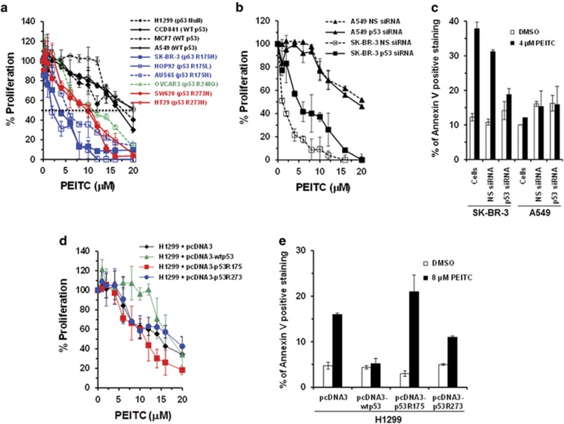
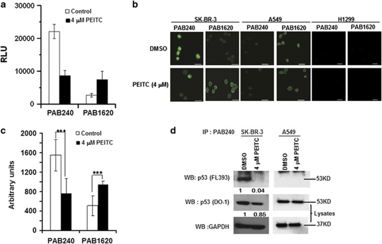
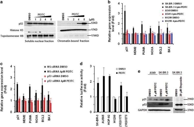
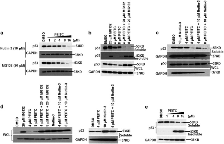
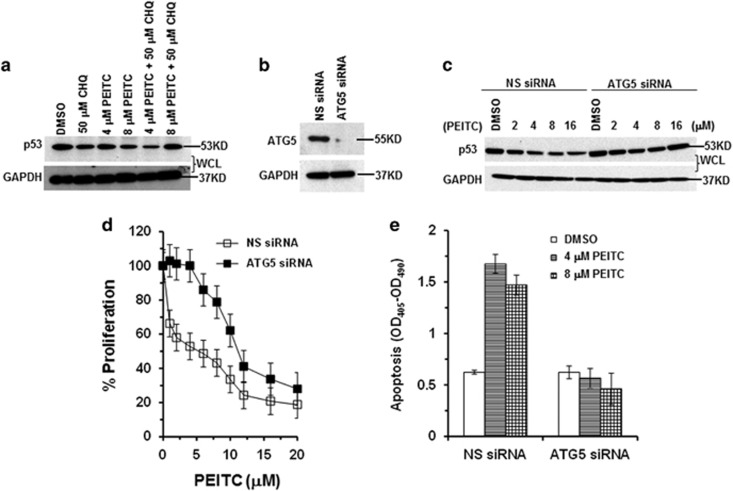
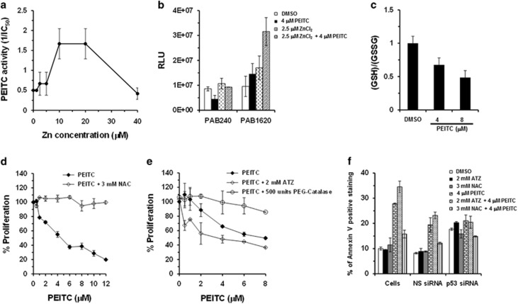
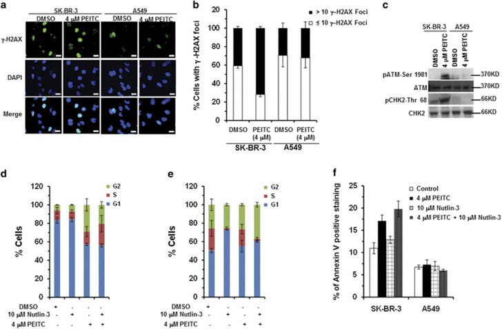
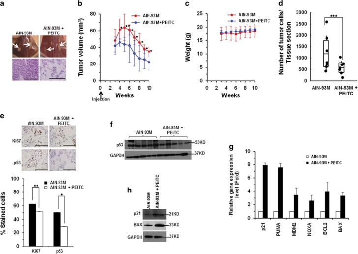
Mutations in the p53 tumor-suppressor gene are prevalent in human cancers. The majority of p53 mutations are missense, which can be classified into contact mutations (that directly disrupts the DNA-binding activity of p53) and structural mutations (that disrupts the conformation of p53). Both of the mutations can disable the normal wild-type (WT) p53 activities. Nevertheless, it has been amply documented that small molecules can rescue activity from mutant p53 by restoring WT tumor-suppressive functions. These compounds hold promise for cancer therapy and have now entered clinical trials. In this study, we show that cruciferous-vegetable-derived phenethyl isothiocyanate (PEITC) can reactivate p53 mutant under in vitro and in vivo conditions, revealing a new mechanism of action for a dietary-related compound. PEITC exhibits growth-inhibitory activity in cells expressing p53 mutants with preferential activity toward p53(R175), one of the most frequent 'hotspot' mutations within the p53 sequence. Mechanistic studies revealed that PEITC induces apoptosis in a p53(R175) mutant-dependent manner by restoring p53 WT conformation and transactivation functions. Accordingly, in PEITC-treated cells the reactivated p53(R175) mutant induces apoptosis by activating canonical WT p53 targets, inducing a delay in S and G2/M phase, and by phosphorylating ATM/CHK2. Interestingly, the growth-inhibitory effects of PEITC depend on the redox state of the cell. Further, PEITC treatments render the p53(R175) mutant sensitive to degradation by the proteasome and autophagy in a concentration-dependent manner. PEITC-induced reactivation of p53(R175) and its subsequent sensitivity to the degradation pathways likely contribute to its anticancer activities. We further show that dietary supplementation of PEITC is able to reactivate WT activity in vivo as well, inhibiting tumor growth in xenograft mouse model. These findings provide the first example of mutant p53 reactivation by a dietary compound and have important implications for cancer prevention and therapy.


Figures

Figure 1
Figure 1
PEITC inhibits cell proliferation and induces apoptosis in a p53R175 mutant-dependent manner. (a) Human tumor cells lines with hotspot p53 mutations and p53 WT were treated with DMSO (control) or PEITC for 3 days. (b) SK-BR-3 and A549 cells transfected with siRNA were treated with DMSO or PEITC for 3 days. Percentage of cell proliferation was determined by the WST-1 assay. (c) Effect of PEITC on apoptosis. Untransfected (cells) or siRNA-transfected SK-BR-3 and A549 cells were treated with DMSO or 4 μM PEITC for 3 days. Apoptosis was measured by Annexin-V staining using a BD LSRFORTESSA instrument. (d) The H1299 cells transfected with pcDNA3, pcDNA3-p53R175, pcDNA3-p53R273 or pcDNA3-wtp53 were treated with DMSO or PEITC for 3 days. Percentage of cell proliferation was determined by the WST-1assay. (e) Effect of PEITC on apoptosis. The H1299 cells transfected with pcDNA3, pcDNA3-p53R175, pcDNA3-p53R273 or pcDNA3-wtp53 were treated with DMSO or 8 μM PEITC for 3 days. Apoptosis was measured by Annexin-V staining using a BD LSRFORTESSA instrument
Figure 2
Figure 2
PEITC induces a ‘WT-like' conformational change in p53R175 mutant protein. (a) ELISA to determine the effect of PEITC on conformation of recombinant-purified GST-p53R175H by using conformation-specific antibodies PAB240 (mutant-specific) and PAB1620 (WT-specific). (b) SK-BR-3 cells were treated with DMSO or 4 μM PEITC for 6 h. Immunofluorescence of the cells was performed using PAB240 and PAB1620 antibodies. The A549 cell line used as a control showed that p53 WT conformation was not changed by PEITC. The H1299 cell line was used as a control for anti-p53 antibodies. All scale bars represents a size of 20 μm. (c) Quantification of PAB240 and PAB1620 staining shown in panel (b). ***P≤0.0001 for PAB240 and PAB1620. (d) Immunoprecipitation of the p53 mutant protein from SK-BR-3 cell lysates using PAB240 antibody and detected by p53 (FL393) antibody
Figure 3
Figure 3
PEITC restores the p53R175 mutant protein transactivational functions. (a) PEITC induced p53R175 mutant protein to bind chromatin. SK-BR-3 cells were treated with PEITC for 4 h and chromatin-bound and nuclear-soluble fractions were analyzed by immunoblotting. Histone H3 and Topoisomerase IIB served as markers for the chromatin and soluble nuclear fractions, respectively. (b) qRT-PCR of p53 regulated genes in SK-BR-3, H1299 and A549 cells treated with DMSO or 4 μM PEITC for 4 h. RNA was extracted and gene expression level was measured using TaqMan gene expression assay. (c) qRT-PCR of p53-regulated genes in NS siRNA or p53 siRNA-transfected SK-BR-3 cells treated with DMSO or 4 μM PEITC for 4 h. RNA was extracted and gene expression level was measured using TaqMan gene expression assay. (d) SK-BR-3, HOP92, AU565, H1299 and MEF ((10)3/175 and (10)3/273) cells were transfected with plasmid 16451 and were treated with PEITC (4 or 6 μM) for 24 h, respectively, followed by a luciferase reporter assay. (e) Western blotting analysis of p21 expression in SK-BR-3 cells treated with 4 μM PEITC or 20 μM etoposide for 4 h. A549 cell line was treated with 4 μM PEITC for 4 h as a control. Protein levels were determined by western blotting using p21, p53 DO-1 and GAPDH antibodies
Figure 4
Figure 4
Proteasome degradation of p53 protein upon PEITC treatment in SK-BR-3 and A549 cells. (a) SK-BR-3 cells were treated with the indicated concentrations of PEITC and inhibitor (10 μM Nutlin-3 or 20 μM MG132) for 4 h. (b) SK-BR-3 cells were treated with PEITC (4 or 8 μM), 20 μM MG132 or both for 4 h. (c) SK-BR-3 cells were treated with PEITC (4 or 8 μM), 10 μM Nutlin-3 or both for 4 h. (d) A549 cells were treated with PEITC (4 or 8 μM), inhibitor (10 μM Nutlin-3 or 20 μM MG132) or both for 4 h. Cells were harvested and lysates were prepared. Lysate fractions were resolved by SDS-PAGE and probed with p53 DO-1 antibody. (e) SK-BR-3 cells were treated with the indicated concentrations of PEITC or DMSO for 4 h. Cells were harvested and soluble and insoluble fractions were prepared. Thirty μg of the soluble and insoluble lysate fractions were resolved by SDS-PAGE and probed with p53 DO-1 antibody
Figure 5
Figure 5
Autophagy of p53R175 protein upon PEITC treatment in SK-BR-3 cells. (a) SK-BR-3 cells were treated with PEITC (4 or 8 μM), CHQ (50 μM) or both for 4 h. Cell lysate fractions were resolved by SDS-PAGE and probed with p53 DO-1 antibody. (b) SK-BR-3 cells were transfected with ATG5 siRNA or NS siRNA. Thirty μg of the cell lysate was resolved by SDS-PAGE and probed with anti-ATG5 antibody. Blots were stripped and reprobed with anti-GAPDH antibody. (c) SK-BR-3 cells transfected with ATG5 siRNA or NS siRNA were treated with DMSO or PEITC for 4 h. Protein levels were determined by western blotting using p53 DO-1 and GAPDH antibodies. (d) SK-BR-3 transfected with ATG5 siRNA or NS siRNA were treated with DMSO or PEITC for 3 days. Percentage of cell proliferation was determined by the WST-1 assay. (e) Effect of PEITC on apoptosis. ATG5 siRNA- or NS siRNA-transfected SK-BR-3 cells were treated with DMSO or PEITC for 3 days. Cells were assayed for histone-associated DNA fragments indicative of apoptosis
Figure 6
Figure 6
Effects of zinc and redox changes on PEITC-induced p53R175 reactivation. (a) Effect of zinc on the activity of PEITC. SK-BR-3 cells were treated with PEITC, zinc or both. Percentage of cell proliferation was determined by the WST-1 assay. The PEITC activity is shown as 1/IC50 for growth inhibition. (b) ELISA to determine the effect of zinc alone or zinc and PEITC on conformation of recombinant-purified GST-p53R175H by using conformation-specific antibodies PAB240 (mutant-specific) and PAB1620 (WT-specific). (c) Effect of PEITC on the levels of reduced glutathione in SK-BR-3 cells. SK-BR-3 cells were treated with PEITC (4 or 8 μM) or DMSO for 4 h. Ratio of reductant GSH and oxidative GSSG was then measured using the GSH/GSSG-Glo Glutathione Assay Kit. (d) Effect of NAC on PEITC activity. SK-BR-3 cells were treated with the indicated concentrations of PEITC or PEITC in combination with 3 mM NAC for 3 days. Percentage of cell proliferation was determined by the WST-1 assay. (e) SK-BR-3 cells were co-treated with the PEITC alone or in combination with 2 mM ATZ or 500 units PEG-Catalase for 3 days. Percentage of cell proliferation was determined by the WST-1 assay. (f) Effect on apoptosis. Untransfected (cells) or siRNA-transfected SK-BR-3 cells were treated with DMSO, ATZ, NAC or PEITC alone or PEITC in combination with ATZ or NAC for 3 days. Apoptosis was measured by Annexin-V staining using a BD LSRFORTESSA instrument
Figure 7
Figure 7
PEITC induces γ-H2AX foci, activates ATM/CHK2, G2/M- and S-phase arrest and apoptosis. SK-BR-3 and A549 cells treated with PEITC or DMSO for 3 days were stained with anti-γ-H2AX antibody. (a) Merged images show cells stained with anti-γ-H2AX antibody (green) and DAPI (blue). All scale bars represents a size of 20 μm. (b) Percentage of cells with γ-H2AX foci (≤10 or >10, as indicated). (c) SK-BR-3 and A549 cells were treated with PEITC or DMSO for 4 h. Western blotting was performed using anti-pATM S1981 and anti-pCHK2 Thr68 antibodies. Blots were stripped and reprobed with anti-ATM and anti-CHK2 antibodies. (d) SK-BR-3 or (e) A549 cells were treated with PEITC, 10 μM Nutlin-3 or both for 24 h and analyzed by flow cytometry. (f) SK-BR-3 and A549 cells were treated with 4μM PEITC, 10 μM Nutlin-3 or both for 24 h. Apoptosis was measured by Annexin-V staining using a BD LSRFORTESSA instrument
Figure 8
Figure 8
PEITC induces p53R175H mutant reactivation in vivo and inhibits xenograft tumor growth. (a) Representative images of mouse mammary fat pads (upper panel), and H&E staining (lower panel). All scale bars represents a size of 200 μm. (b) Tumors were measured with Vernier calipers, and tumor volumes were calculated. Formula L × W2 × 0.523 (**P≤0.009 and *P≤0.03; n=7). (c) Animal weights (g) were measured weekly. (d) Distribution of the animals based on the average number of tumor cells per tissue section in the control and PEITC groups (***P≤0.00026; n=7). (e) Representative images of xenograft tumor tissue stained for Ki67 (**P≤0.007) and p53 (*P≤0.033) (upper panel) and quantitation of positive cells (lower panel) (n=7). Results are expressed as ±S.D. All scale bars represents a size of 200 μm. (f) Western blotting analysis of p53 expression levels in the xenograft tumors from the PEITC and control animal groups. Blot is representative of the 12 tumor tissue lysates analyzed from each group. (g) qRT-PCR (n=4) of p53-regulated genes in the PEITC and control animal groups. (h) Western blotting of p21 and Bax expression in SK-BR-3 xenograft tumors in vivo

References

    1. Liu G, McDonnell TJ, Montes de Oca Luna R, Kapoor M, Mims B, El-Naggar AK et al. High metastatic potential in mice inheriting a targeted p53 missense mutation. Proc Natl Acad Sci USA 2000; 97: 4174–4179. - PMC - PubMed
    1. Muller PA, Caswell PT, Doyle B, Iwanicki MP, Tan EH, Karim S et al. Mutant p53 drives invasion by promoting integrin recycling. Cell 2009; 139: 1327–1341. - PubMed
    1. Dittmer D, Pati S, Zambetti G, Chu S, Teresky AK, Moore M et al. Gain of function mutations in p53. Nat Genet 1993; 4: 42–46. - PubMed
    1. Hisada M, Garber JE, Fung CY, Fraumeni JF Jr, Li FP. Multiple primary cancers in families with Li-Fraumeni syndrome. J Natl Cancer Inst 1998; 90: 606–611. - PubMed
    1. WHOIARC Handbook on Cancer Prevention. Cruciferous Vegetables, Isothiocyanates and Indoles, vol. 9. IARC Press: Lyon, France, 2004.

MeSH terms