. 2016 Jun 2;98(6):1193-1207.
doi: 10.1016/j.ajhg.2016.05.004.

Mutations in GANAB, Encoding the Glucosidase IIα Subunit, Cause Autosomal-Dominant Polycystic Kidney and Liver Disease

Affiliations

Mutations in GANAB, Encoding the Glucosidase IIα Subunit, Cause Autosomal-Dominant Polycystic Kidney and Liver Disease

Binu Porath et al. Am J Hum Genet. .

Abstract

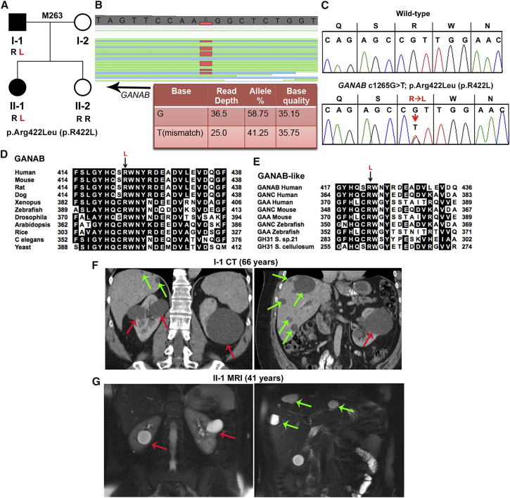
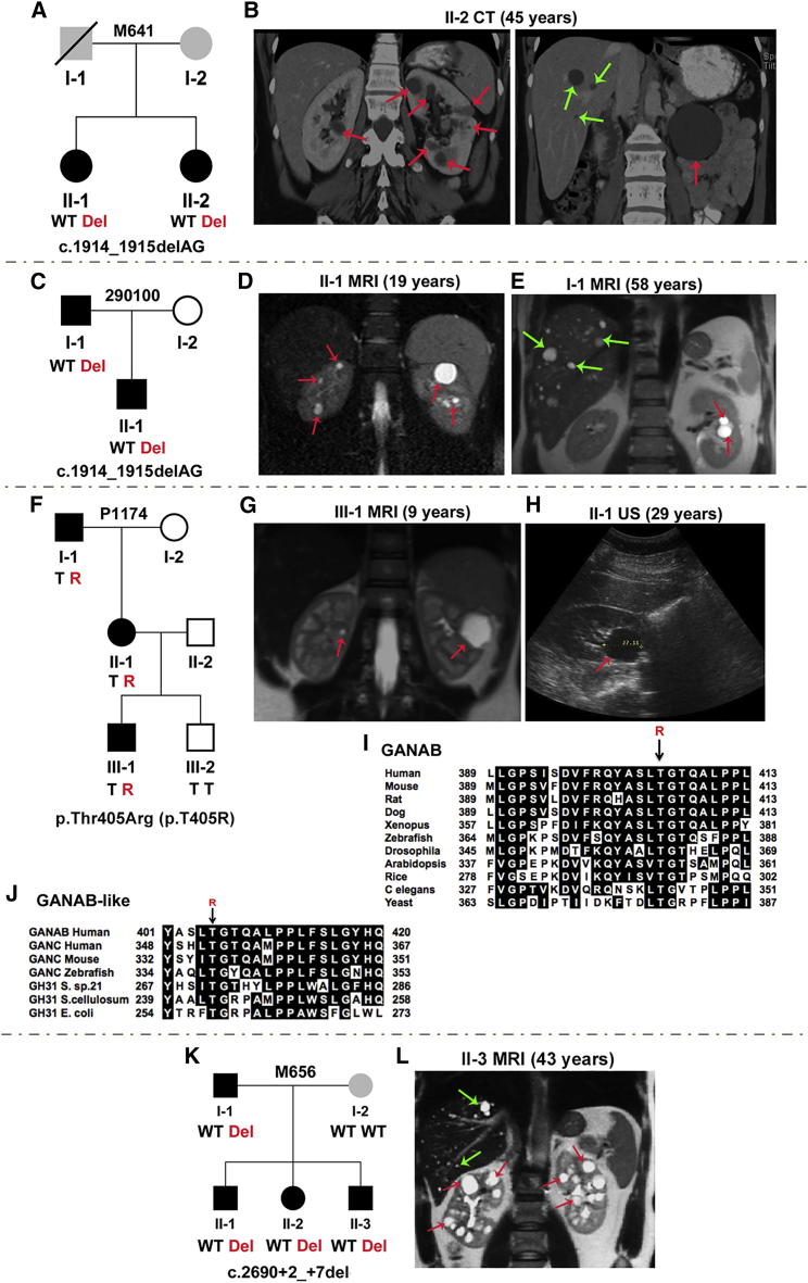
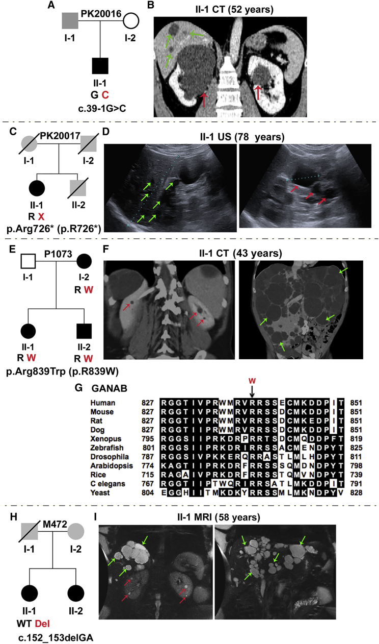
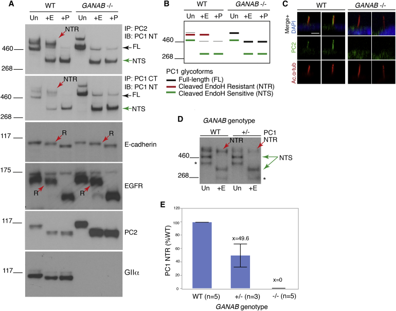
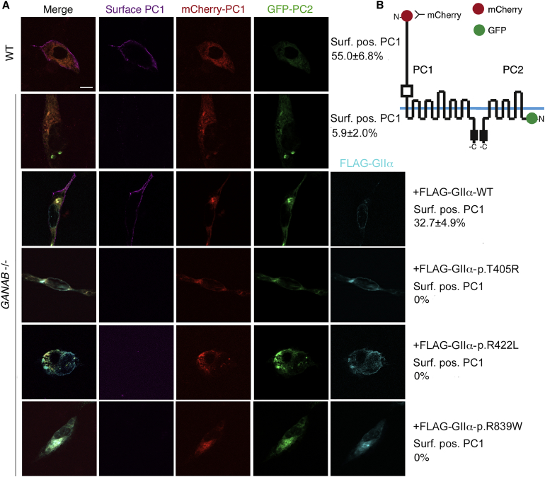
Autosomal-dominant polycystic kidney disease (ADPKD) is a common, progressive, adult-onset disease that is an important cause of end-stage renal disease (ESRD), which requires transplantation or dialysis. Mutations in PKD1 or PKD2 (∼85% and ∼15% of resolved cases, respectively) are the known causes of ADPKD. Extrarenal manifestations include an increased level of intracranial aneurysms and polycystic liver disease (PLD), which can be severe and associated with significant morbidity. Autosomal-dominant PLD (ADPLD) with no or very few renal cysts is a separate disorder caused by PRKCSH, SEC63, or LRP5 mutations. After screening, 7%-10% of ADPKD-affected and ∼50% of ADPLD-affected families were genetically unresolved (GUR), suggesting further genetic heterogeneity of both disorders. Whole-exome sequencing of six GUR ADPKD-affected families identified one with a missense mutation in GANAB, encoding glucosidase II subunit α (GIIα). Because PRKCSH encodes GIIβ, GANAB is a strong ADPKD and ADPLD candidate gene. Sanger screening of 321 additional GUR families identified eight further likely mutations (six truncating), and a total of 20 affected individuals were identified in seven ADPKD- and two ADPLD-affected families. The phenotype was mild PKD and variable, including severe, PLD. Analysis of GANAB-null cells showed an absolute requirement of GIIα for maturation and surface and ciliary localization of the ADPKD proteins (PC1 and PC2), and reduced mature PC1 was seen in GANAB(+/-) cells. PC1 surface localization in GANAB(-/-) cells was rescued by wild-type, but not mutant, GIIα. Overall, we show that GANAB mutations cause ADPKD and ADPLD and that the cystogenesis is most likely driven by defects in PC1 maturation.


Figures

Figure 1
Figure 1
WES Analysis Reveals GIIα p.Arg422Leu (c.1265G>T) in Family M263 as the Likely Pathogenic Variant (A) Pedigree of family M263 shows that the two affected individuals (I-1 and II-1, shaded in black) have the GIIα p.Arg422Leu (p.R422L) missense variant resulting from c.1256G>T in exon 12, but the unaffected daughter (II-2, for whom no cysts were detected on ultrasound at 30 years of age) does not. (B) GenomeBrowse (SVS, Golden Helix) view of the WES results from II-1 show GANAB variant c.1265G>T (reverse strand), and details of the reads are tabulated below. (C) Sanger sequencing confirmation of heterozygous GIIα variant p.Arg422Leu (p.R422L) (c.1256G>T) in II-1. The WT is shown for comparison. (D) MSA of GIIα orthologous proteins shows invariance of Arg422 from humans to yeast. In silico analysis of the likely pathogenicity of GIIα p.Arg422Leu (p.R422L) shows variant scores (SIFT = 0.00, Align GVGD = C65) characteristic of a highly likely pathogenic mutation. (E) MSA of related glucosidases GANC (neutral alpha-glucosidase C) and GAA (lysosomal alpha-glucosidase) of various eukaryotic species and prokaryotic GH31 (glycosyl hydrolase family 31) shows invariant conservation of GIIα Arg422. (F) CT scan with contrast of kidneys and liver of individual I-1 at 66 years shows a few large kidney cysts (red arrows) and multiple scattered liver cysts (green arrows). (G) T2-weighted MRI of II-1 at 41 years shows a few kidney (red arrows) and liver (green arrows) cysts.
Figure 2
Figure 2
Characterization of GANAB Mutations in Four ADPKD-Affected Families (A) Pedigree of family M641 shows c.1914_1915delAG (p.Asp640Glnfs77) (exon 17) in two affected siblings. Both sisters had basilar tip aneurysms, and II-2 also had two aneurysms detected on the left middle cerebral artery. The affected status of the parents is unclear (gray shading); I-1 had renal cell carcinoma and a ruptured aneurysm, but no reported PKD. (B) CT scan of kidneys and liver from II-2 shows multiple kidney and occasional liver cysts. (C) Pedigree of family 290100 shows c.1914_1915delAG in the father and son. (D and E) MRI shows a few kidney but no hepatic cysts in II-1 (D) and a few kidney and scattered liver cysts in I-1 (E). (F) Pedigree of family P1174 shows p.Thr405Arg (p.T405R) (c.1214C>G, exon 11) in three affected individuals; III-2, for whom no cysts were detected by ultrasound at 5 years, did not have the variant. (G and H) MRI of III-1 shows bilateral kidney cysts (G), and ultrasound (US) of II-1 shows a single large renal cyst (H). (I) MSA of GIIα orthologs shows that Thr405 is invariant across species. In silico mutation analysis highly predicts p.Thr405Arg (p.T405R) to be pathogenic (SIFT = 0.00, Align GVGD = C65). (J) MSA of GANAB-like proteins shows invariant conservation of this residue. (K) Pedigree of family M656 shows c.2690+2_+7del (IVS20) in four affected members. Splicing predictions show complete loss of the donor site. The mother (I-2), who does not have the GANAB mutation, has ∼5 kidney and ∼30 liver cysts, but no detected PKD1, PKD2, PRKCSH, or SEC63 mutations. (L) MRI of II-3 shows small hepatic andmultiple renal cysts. Red and green arrows indicate kidney and liver cysts, respectively. Where multiple cysts are present, only representative cysts are highlighted.
Figure 3
Figure 3
Characterization of GANAB Variants in Four Families, Including ADPLD-Affected P1073 and M472 (A) Pedigree of family PK20016 shows the splicing mutation c.39−1G>C (IVS1). The family history is unclear because samples were unavailable, but one large renal cyst was reported in I-1. (B) CT scan of II-1 shows bilateral kidney cysts and occasional hepatic cysts. (C) Pedigree of family PK20017 shows p.Arg726 (p.R726) (c.2176C>T, exon 18), in the proband, II-1. A lack of DNA samples and clinical information precluded determining the family history. II-2 died at 55 years from a ruptured intracranial aneurysm, but his PKD status was unknown. (D) Ultrasound examination of II-1 shows several liver (left), and kidney cysts (right). (E) Pedigree of family P1073 shows p.Arg839Trp (p.R839W) (c.2515C>T, exon 22) in three affected individuals. (F) CT scan of II-1 shows very few kidney cysts (left) but severe PLD (right). Gross images of the liver of this subject have been published. (G) MSA of GANAB (GIIα) orthologs shows invariant conservation of Arg839 across species. In silico mutation analysis highly predicts p.Arg839Trp (p.R839W) to be pathogenic (SIFT = 0.00, Align GVGD = C65). (H) Pedigree of family M472 shows c.152_153delGA (p.Arg51Lysfs21) (p.R51fs; exon 3) in II-1. (I) MRI of II-1 shows a few renal cysts (left) but significant PLD; this image was subsequent to earlier resections (Table 1). No sample was available from II-2, but imaging also showed predominant PLD (Figure S3E). Unavailable parental DNA samples and limited clinical information precluded determining the family history, but I-1 was reported to have had a cerebral hemorrhage. Red and green arrows indicate kidney and liver cysts, respectively. Where multiple cysts are present, only representative cysts are highlighted.
Figure 4
Figure 4
GIIα Is Required for PC1 ER Exit and Maturation (A) Deglycosylation analysis of WT and GANAB−/− RCTE membrane protein either untreated (Un) or treated with EndoH (+E) or PNGaseF (+P). IP was used to enrich the PC1 complex with C-terminal PC1 (PC1-CT) or PC2 (YCE2) antibodies and immunodetected with the N-terminal PC1 (PC1-NT) antibody (7e12). Complete loss of the mature PC1 glycoform (NTR; red arrow) was observed in GANAB−/− cells, and full-length PC1 (FL) and PC1-NTS became more abundant. Mature E-cadherin and EGFR were not or only marginally affected by GANAB loss; the EndoH-resistant protein (R, red arrow) persisted. No EndoH-resistant form of PC2 was noted, but the protein was upregulated in GANAB−/− cells. Loss of WT GIIα was confirmed in GANAB−/− cells with the use of a C-terminal antibody. (B) Schematic representation of the observed PC1 banding pattern in the WT and GANAB−/− cells shown in (A). (C) Confocal z stack rendering of primary cilia in confluent WT and GANAB−/− cells in which acetylated α-tubulin (Ac.α-tub) and PC2 were detected shows no cilia PC2 signal in GANAB−/− cells. Nuclei were stained with DAPI, and 100 ciliated cells were analyzed in three independent experiments. The scale bar represents 10 μm. (D) Immunoblot of PC1-NT in WT and GANAB+/− cells shows a reduced level of PC1-NTR in GANAB+/− cells. Asterisks indicate a non-specific product. (E) Quantification of PC1-NTR shows a reduction to ∼50% (p < 0.001, Student’s t test) in heterozygous cells and a complete loss in homozygous, GANAB−/− cells.
Figure 5
Figure 5
Surface Localization of PC1 Requires WT GIIα and Is Disrupted by GANAB Missense Mutations (A) WT and GANAB−/− cells were co-transfected with WT tagged PC1 and PC2, mCherry-PC1, and TagGFP-PC2 and examined for surface mCherry-PC1 labeling. Co-transfected cells were screened for live cell-surface PC1 signal and quantified as the percentage of surface-positive PC1 cells out of the total co-transfected cells. Surface PC1 was detected on 55.0% ± 6.8% of WT cells but only 5.9% ± 2.0% of GANAB−/− cells (p ≤ 0.0001), and the level was rescued to 32.7% ± 4.9% by co-transfection with the WT GANAB (FLAG-GIIα) plasmid. Co-transfection with the newly identified putative GANAB missense mutations cloned in FLAG-GIIα, p.Thr405Arg (p.T405R), p.Arg422Leu (p.R422L), and p.Arg839Trp (p.R839W) did not rescue PC1 surface localization (bottom three panels; all p < 0.0001 versus WT rescue). Student’s t test was performed to determine significance in at least 100 triple-transfected cells analyzed among three independent experiments. The scale bar represents 20 μm. (B) Diagram of the constructs.

References

    1. Torres V.E., Harris P.C., Pirson Y. Autosomal dominant polycystic kidney disease. Lancet. 2007;369:1287–1301. - PubMed
    1. Harris P.C., Torres V.E. Polycystic kidney disease. Annu. Rev. Med. 2009;60:321–337. - PMC - PubMed
    1. The European Polycystic Kidney Disease Consortium The polycystic kidney disease 1 gene encodes a 14 kb transcript and lies within a duplicated region on chromosome 16. Cell. 1994;77:881–894. - PubMed
    1. Mochizuki T., Wu G., Hayashi T., Xenophontos S.L., Veldhuisen B., Saris J.J., Reynolds D.M., Cai Y., Gabow P.A., Pierides A. PKD2, a gene for polycystic kidney disease that encodes an integral membrane protein. Science. 1996;272:1339–1342. - PubMed
    1. Heyer C.M., Sundsbak J.L., Abebe K.Z., Chapman A.B., Torres V.E., Grantham J.J., Bae K.T., Schrier R.W., Perrone R.D., Braun W.E., HALT PKD and CRISP Investigators Predicted mutation strength of nontruncating PKD1 mutations aids genotype-phenotype correlations in autosomal dominant polycystic kidney disease. J. Am. Soc. Nephrol. 2016 Published online January 28, 2016. - PMC - PubMed

Publication types

MeSH terms

Substances

Supplementary concepts