. 2016 Jun 2;165(6):1440-1453.
doi: 10.1016/j.cell.2016.05.037.

Dual RING E3 Architectures Regulate Multiubiquitination and Ubiquitin Chain Elongation by APC/C

Affiliations

Dual RING E3 Architectures Regulate Multiubiquitination and Ubiquitin Chain Elongation by APC/C

Nicholas G Brown et al. Cell. .

Abstract

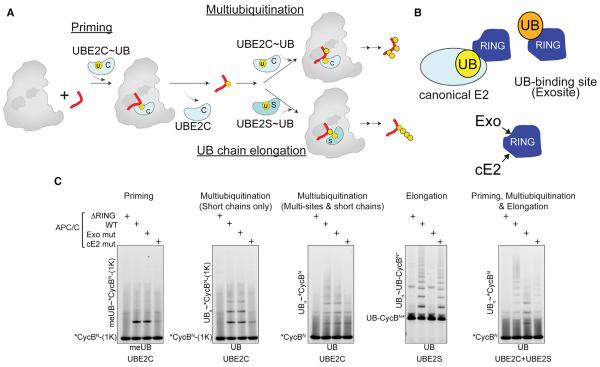
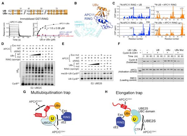
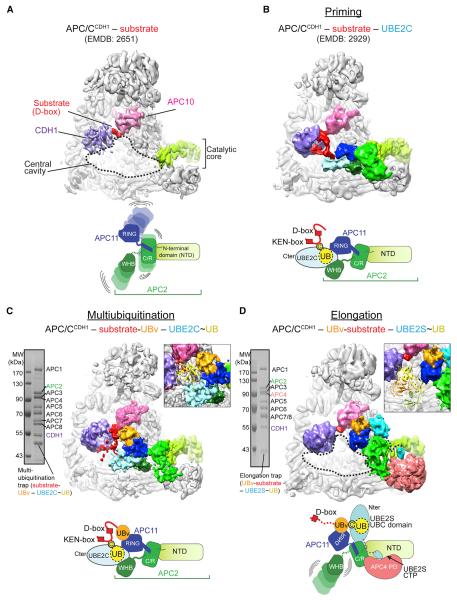
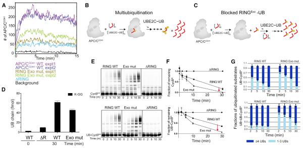
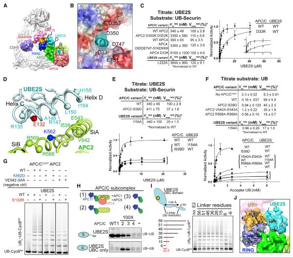
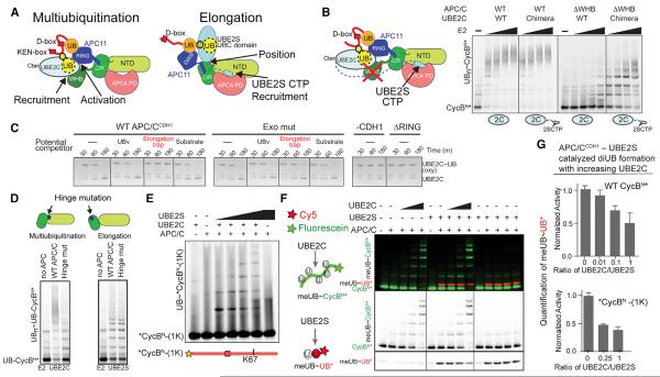
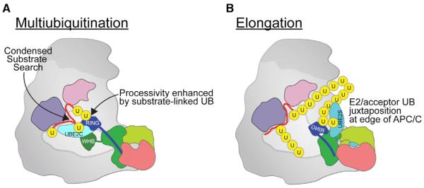
Protein ubiquitination involves E1, E2, and E3 trienzyme cascades. E2 and RING E3 enzymes often collaborate to first prime a substrate with a single ubiquitin (UB) and then achieve different forms of polyubiquitination: multiubiquitination of several sites and elongation of linkage-specific UB chains. Here, cryo-EM and biochemistry show that the human E3 anaphase-promoting complex/cyclosome (APC/C) and its two partner E2s, UBE2C (aka UBCH10) and UBE2S, adopt specialized catalytic architectures for these two distinct forms of polyubiquitination. The APC/C RING constrains UBE2C proximal to a substrate and simultaneously binds a substrate-linked UB to drive processive multiubiquitination. Alternatively, during UB chain elongation, the RING does not bind UBE2S but rather lures an evolving substrate-linked UB to UBE2S positioned through a cullin interaction to generate a Lys11-linked chain. Our findings define mechanisms of APC/C regulation, and establish principles by which specialized E3-E2-substrate-UB architectures control different forms of polyubiquitination.


Figures

Figure 1
Figure 1. APC/C and Two E2 Partners Use Distinct Mechanisms for Polyubiquitination
(A) Distinct mechanisms of priming, multiubiquitination and UB chain elongation by APC/C, UBE2C, and UBE2S. (B) Distinct APC11 RING surfaces involved in polyubiquitination: canonical E2 binding site - cE2; UB-binding exosite - Exo. (C) APC11 RING mutants define distinctive priming, multiubiquitination, and UB chain elongation by APC/CCDH1 with UBE2C and/or UBE2S, using WT or methyl UB (meUB), during a single encounter of the indicated version of CycBN (1K = K67 only; UB- = UB-fusion).
Figure 2
Figure 2. Anchoring the RINGexo Site, UBE2C, and UBE2S for Structural Studies of Polyubiquitination
(A) Phage display selected UB variant (UBv) binds APC11 RING with high affinity and selectivity, measured by BLI. (B) Crystal structure UBv (orange)–APC11 (navy) confirms binding to exosite, opposite canonical E2 site (modeled, cyan). (C) UB and UBv bind similar APC11 RING surface and vice-versa, based on NMR chemical shift perturbations (CSPs). (D) Similar effects on UBE2C-dependent multiubiquitination for blocking RING exosite with either UBv or APC11 mutations. Sponge = excess APC11 RING sequestering UBv. (E) Similar effects on UBE2S-dependent UB chain elongation for blocking RING exosite with either UBv or APC11 mutations. (F) UBv inhibits APC/C-dependent degradation of cyclin B in mitotic X. laevis egg extracts, examined by indicated westerns. (G) Scheme of 3-way crosslinked complex used to trap APC/CCDH1–UBE2C architecture representing multiubiquitination. (H) Scheme of 3-way crosslinked complex used to trap APC/CCDH1–UBE2S architecture representing UB chain elongation.
Figure 3
Figure 3. “Snapshots” of Distinct APC/C–E2 Architectures for Polyubiquitination
(A) Prior APC/CCDH1–substrate complex (Chang et al., 2014). CDH1-purple, APC10-pink, APC2 NTD-light green, substrate-red. (B) Prior structural data for complex representing priming (Brown et al., 2015), with UBE2C in light blue, APC2–APC11 intermolecular cullin–RING (C/R) domain green, and APC2 WHB domain in forest. (C) Cryo-EM reconstruction representing multiubiquitination. UBv - orange. Inset, one UB (1UBQ, yellow) is shown fitting between substrate and active site. (D) Cryo-EM reconstruction representing UB chain elongation. UBE2S - teal. Inset, distance between substrate binding and active sites accommodates polyUB, shown by tetraUB (2XEW, cartoon).
Figure 4
Figure 4. Substrate-Linked UB Binding to RING Exosite Contributes to Processive Affinity Amplification for Multiubiquitination
(A) Single-molecule time traces for binding to evolving ubiquitinated immobilized Securin molecules during multiubiquitination by UBE2C and APC/CCDH1 or indicated RING mutants. (B) In processive multiubiquitination by APC/C and UBE2C, multiple UBs are added to substrate in a single binding event. (C) Model if blocking substrate-linked UB binding to the RING exosite reduces processivity and shifts to distributive mode of multiubiquitination. A larger fraction of substrate would be modified, but with fewer UBs. (D) RING exosite contributes to quantity of UB chains formed during CycBN* multiubiquitination by APC/CCDH1 and UBE2C, measured by AQUA mass spectrometry. (E) Role of UB-binding RING exosite on processivity, monitored by formation of high molecular weight conjugates and fraction of substrate modified during multiple turnover UBE2C-catalyzed multiubiquitination of CycBN* (top) or UB-CycBN* (bottom). (F) Role of RING exosite on fraction of substrate modified over time, in assays as in (E), quantifying depletion of unmodified CycBN* (top) or UB-CycBN* (bottom). Error bars: SEM, N = 3. (G) Role of RING exosite on extent of substrate modification during multiubiquitination with UBE2C. Ubiquitinated products generated as in (E) were divided into 2 categories, with ≥ 4 UBs (navy) or 1–3 UBs (blue) as resolved by SDS-PAGE to examine extent of generation of highly multiubiquitinated products. Error bars: SD, N = 3.
Figure 5
Figure 5. Distinctive Multisite Interactions Establish Unique Catalytic Architecture Specifying UB Chain Elongation by APC/C and UBE2S
(A) Cryo-EM reconstruction of APC/C-CDH1 complex with UBE2S representing UB chain elongation as in Figure 3D, indicating regions with close-ups in panels (B), (D), and (J). (B) Model for APC2/APC4 groove interactions with UBE2S CTP, based on docking APC/C structure (Chang et al., 2015) in cryo-EM reconstruction. APC2/APC4 groove is shown as a surface colored by electrostatic potential, with selected side-chains lining the groove in spheres. EM density for UBE2S CTP - cyan. (C) Role of APC2/APC4 groove in recruiting UBE2S CTP, as determined from kinetic parameters for the indicated mutants during polyubiquitination of a UBSecurin substrate while titrating UBE2S. SEM, n ≥ 3. (D) Placement of UBE2S C- and D-helices (cyan) by APC2 Si helices modeled based on (Chang et al., 2015) (green). (E) Role of APC2 placement of UBE2S UBC domain in substrate polyubiquitination, from kinetic parameters for mutants during polyubiquitination of a UB-Securin substrate while titrating UBE2S. SEM, n ≥ 3. (F) Role of APC2 placement of UBE2S UBC domain in activating UB chain synthesis, from kinetic parameters for mutants upon titrating acceptor UB during APC/CCDH1-UBE2S-mediated di-UB synthesis. SEM, n ≥ 3. (G) APC2 placement of UBE2S UBC domain tested by charge-swap rescue assaying UBE2S E132R restoring UB chain elongation specifically to APC/CCDH1 with APC2 K562D mutant. (H) Importance of placing UBE2S's UBC domain, or recruiting the CTP, determined from minimal APC/C subcomplexes (schematics on top) required to stimulate di-UB synthesis by WT UBE2S or isolated UBC domain lacking the CTP (bottom). Reactions with APC/C WT and subcomplexes 1–3 with WT UBE2S are controls based on (Brown et al., 2014). (I) Importance of placing both UBE2S's UBC domain and CTP for APC/C activation of UB chain elongation. UBE2S deletion mutants with progressively shorter linkers between the two domains were assayed for APC/CCDH1-dependent polyubiquitination of UB-CycBN*. Reactions with WT UBE2S and linker deletions to 26 are controls based on (Brown et al., 2014). (J) Model for acceptor UB (orange, UBv as proxy) in active site of UBE2S (cyan).
Figure 6
Figure 6. Functional Specialization of Each Polyubiquitination Architecture
(A) Distinct APC/C mechanisms recruiting, positioning, and/or activating UBE2C or UBE2S for multiubiquitination and UB chain elongation, respectively. (B) UBE2S CTP is a poor substitute for APC2 WHB in supporting UBE2C-dependent substrate multiubiquitination, as shown from assays with WT UBE2C or a chimera with appended UBE2S's CTP, and WT APC/CCDH1 or a deletion mutant lacking APC2 WHB domain. (C) Shackling the RING away from the multiubiquitination architecture by the elongation trap inhibits APC/C activation of intrinsic UBE2C activity, monitored by inhibition of APC/CCDH1-stimulated hydrolysis of oxyester-linked UBE2C~UB. (D) Specific APC2 cullin conformation is required for multiubiquitination. Top – schematic of distinctive APC2 NTD-C/R domain orientations showing burial or exposure of hinge. Distinct defects with hinge mutant (APC2 I501D, I502D) for multiubiquitination or UB chain elongation of UB-CycBN*. (E) UBE2C and UBE2S build a UB chain on *CycBN-(1K) during the substrate's single encounter with APC/CCDH1. (F) Competition between APC/CCDH1 activities with UBE2C and UBE2S probed simultaneously with 2 colors. MeUB can only be donor and not acceptor. Only fluorescein-CycBN* (green) accepts meUB from UBE2C. Only Cy5-UB* (red) with blocked C terminus accepts meUB from UBE2S. (G) Bar graphs showing reduced APC/CCDH1-UBE2S-catalyzed meUB~UB* formation in (F) and S6G in the presence of UBE2C activity. SD, n ≥ 2.
Figure 7
Figure 7. Specialized APC/C–E2 Architectures for Multiubiquitination and UB Chain Elongation
(A) Processive multiubiquitination occurs by APC/C's APC2 cullin (green)-APC11 RING (blue) positioning UBE2C proximal to substrate (red), reducing the search volume for catalytic encounter while substrate-linked UB (yellow) binds the RING exosite to increase the evolving ubiquitinated substrate's lifetime on APC/C and enhance processivity. Each UB transfer cycle is accompanied by catalytic core dynamics releasing the used UBE2C for replacement by a charged UBE2C~UB to donate the next UB for ligation. (B) Specialized architecture for UB chain elongation. APC2/APC4 recruits UBE2S's CTP, APC2 (cullin) places UBE2S's catalytic UBC domain, and APC11's RING guides the acceptor UB's Lys11 to the active site. Location of UBE2S at the edge of APC/C would accommodate growth of long UB chains.

References

    1. Aristarkhov A, Eytan E, Moghe A, Admon A, Hershko A, Ruderman JV. E2-C, a cyclin-selective ubiquitin carrier protein required for the destruction of mitotic cyclins. Proc. Natl. Acad. Sci. USA. 1996;93:4294–4299. - PMC - PubMed
    1. Berndsen CE, Wolberger C. New insights into ubiquitin E3 ligase mechanism. Nat. Struct. Mol. Biol. 2014;21:301–307. - PubMed
    1. Branigan E, Plechanovová A, Jaffray EG, Naismith JH, Hay RT. Structural basis for the RING-catalyzed synthesis of K63-linked ubiquitin chains. Nat. Struct. Mol. Biol. 2015;22:597–602. - PMC - PubMed
    1. Brown NG, Watson ER, Weissmann F, Jarvis MA, VanderLinden R, Grace CR, Frye JJ, Qiao R, Dube P, Petzold G, et al. Mechanism of polyubiquitination by human anaphase-promoting complex: RING repurposing for ubiquitin chain assembly. Mol. Cell. 2014;56:246–260. - PMC - PubMed
    1. Brown NG, VanderLinden R, Watson ER, Qiao R, Grace CR, Yamaguchi M, Weissmann F, Frye JJ, Dube P, Ei Cho S, et al. RING E3 mechanism for ubiquitin ligation to a disordered substrate visualized for human anaphase-promoting complex. Proc. Natl. Acad. Sci. USA. 2015;112:5272–5279. - PMC - PubMed

Publication types

MeSH terms

Substances