

The novel in vitro reanimation of isolated human and large mammalian heart-lung blocs
- PMID: 27259478
- PMCID: PMC4893289
- DOI: 10.1186/s12899-016-0023-2
The novel in vitro reanimation of isolated human and large mammalian heart-lung blocs
Abstract
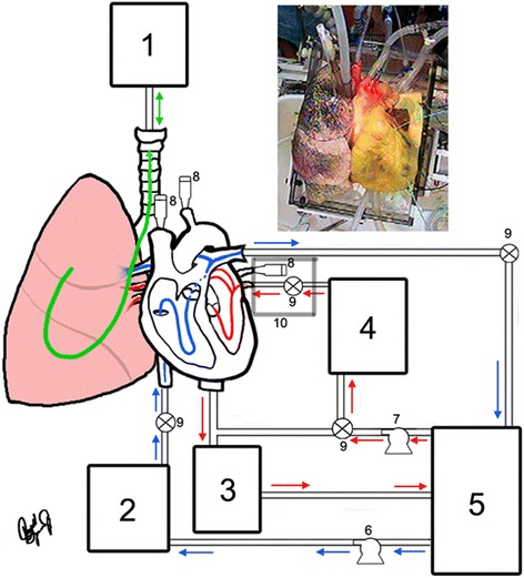
Background: In vitro isolated heart preparations are valuable tools for the study of cardiac anatomy and physiology, as well as for preclinical device testing. Such preparations afford investigators a high level of hemodynamic control, independent of host or systemic interactions. Here we hypothesize that recovered human and swine heart-lung blocs can be reanimated using a clear perfusate and elicit viable cardiodynamic and pulmonic function. Further, this approach will facilitate multimodal imaging, which is particularly valuable for the study of both functional anatomy and device-tissue interactions. Five human and 18 swine heart-lung preparations were procured using techniques analogous to those for cardiac transplant. Specimens were then rewarmed and reperfused using modifications of a closed circuit, isolated, beating and ventilated heart-lung preparation. Positive pressure mechanical ventilation was also employed, and epicardial defibrillation was applied to elicit native cardiac sinus rhythm. Videoscopy, fluoroscopy, ultrasound, and infrared imaging were performed for anatomical and experimental study.
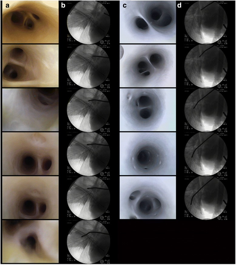
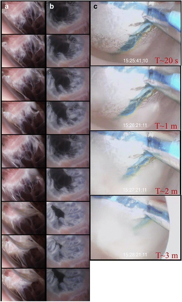
Results: Systolic and diastolic left ventricular pressures observed for human and swine specimens were 68/2 ± 11/7 and 74/3 ± 17/5 mmHg, respectively, with associated native heart rates of 80 ± 7 and 96 ± 16 beats per minute. High-resolution imaging within functioning human pulmonary vasculature was obtained among other anatomies of interest. Note that one human specimen elicited poor cardiac performance post defibrillation.
Conclusions: We report the first dynamic videoscopic images of the pulmonary vasculature during viable cardiopulmonary function in isolated reanimated heart-lung blocs. This experimental approach provides unique in vitro opportunities for the study of novel medical therapeutics applied to large mammalian, including human, heart-lung specimens.
Keywords: Cardiac anatomy; Cardiac physiology; Heart-lung bloc; Reanimation.
Figures
Similar articles
-
In vitro studies of human hearts.Ann Thorac Surg. 2005 Jan;79(1):168-77. doi: 10.1016/j.athoracsur.2004.06.080. Ann Thorac Surg. 2005. PMID: 15620938
-
In vivo versus in vitro comparison of swine cardiac performance: induction of cardiodepression with halothane.Eur J Pharmacol. 2006 Aug 14;543(1-3):97-107. doi: 10.1016/j.ejphar.2006.06.011. Epub 2006 Jun 10. Eur J Pharmacol. 2006. PMID: 16842774
-
Multimodal functional and still imaging of a transplanted human heart reanimated using Visible Heart® methodologies.J Card Surg. 2020 Mar;35(3):668-671. doi: 10.1111/jocs.14403. Epub 2020 Jan 16. J Card Surg. 2020. PMID: 31945224
-
Interactions: the integrated functioning of heart and lungs.Adv Exp Med Biol. 1993;346:347-64. doi: 10.1007/978-1-4615-2946-0_34. Adv Exp Med Biol. 1993. PMID: 8184775 Review.
-
The benefits of the Atlas of Human Cardiac Anatomy website for the design of cardiac devices.Expert Rev Med Devices. 2013 Nov;10(6):729-34. doi: 10.1586/17434440.2013.843449. Expert Rev Med Devices. 2013. PMID: 24195457 Review.
Cited by
-
Effects of ATP administration on isolated swine hearts: Implications for ex vivo perfusion and cardiac transplantation.Exp Biol Med (Maywood). 2019 Aug;244(11):915-922. doi: 10.1177/1535370219850786. Epub 2019 May 27. Exp Biol Med (Maywood). 2019. PMID: 31132883 Free PMC article.
References
-
- Langendorff O. Untersuchungen am überlebenden säugethierherzen. Arch Für Gesamte Physiol Menschen Tiere. 1895;61:291–332. doi: 10.1007/BF01812150. - DOI
MeSH terms
LinkOut - more resources
Full Text Sources
Other Literature Sources
