Role of Klotho in migration and proliferation of human dermal microvascular endothelial cells
- PMID: 27260080
- PMCID: PMC4938740
- DOI: 10.1016/j.mvr.2016.05.005
Role of Klotho in migration and proliferation of human dermal microvascular endothelial cells
Abstract
Purpose: To examine the possible role of Klotho (Kl) in human microvasculature.
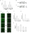
Methods: The expression level of Kl in primary human dermal microvascular endothelial cells (HDMECs) and primary human dermal fibroblasts (HFb) was detected by real-time polymerase chain reaction amplification (qRT-PCR), Western blot analyses and immunohistochemistry. Migration of HDMECs and HFb was examined in monolayer wound healing "scratch assay" and Transwell assay. Proliferation of these cells was examined using Cell Proliferation BrdU incorporation assay.
Results: Our results have shown that downregulation of Kl abrogated HDMECs migration after 48h. On the other hand, migration of HFb significantly increased after blocking Kl. Lack of Kl decreased expression of genes involved in the activation of endothelial cells and enhanced expression of genes involved in extracellular matrix remodeling and organization of connective tissue.
Conclusions: This study for the first time provides the evidence that Kl is expressed in HDMECs and HFb. Additionally, we have demonstrated that Kl is implicated in the process of angiogenesis of human dermal microvasculature.
Keywords: Angiogenesis; Dermal fibroblasts; Dermal microvascular endothelial cells; Klotho.
Published by Elsevier Inc.
Figures


Similar articles
-
Proangiogenic effects of soluble α-Klotho on systemic sclerosis dermal microvascular endothelial cells.Arthritis Res Ther. 2017 Feb 10;19(1):27. doi: 10.1186/s13075-017-1233-0. Arthritis Res Ther. 2017. PMID: 28183357 Free PMC article.
-
A new in vitro wound model based on the co-culture of human dermal microvascular endothelial cells and human dermal fibroblasts.Biol Cell. 2007 Apr;99(4):197-207. doi: 10.1042/BC20060116. Biol Cell. 2007. PMID: 17222082
-
α-Klotho expression determines nitric oxide synthesis in response to FGF-23 in human aortic endothelial cells.PLoS One. 2017 May 2;12(5):e0176817. doi: 10.1371/journal.pone.0176817. eCollection 2017. PLoS One. 2017. PMID: 28463984 Free PMC article.
-
Increased angiogenesis and migration of dermal microvascular endothelial cells from patients with psoriasis.Exp Dermatol. 2021 Jul;30(7):973-981. doi: 10.1111/exd.14329. Epub 2021 Mar 22. Exp Dermatol. 2021. PMID: 33751661
-
Abnormal dermal microvascular endothelial cells in psoriatic excessive angiogenesis.Microvasc Res. 2024 Sep;155:104718. doi: 10.1016/j.mvr.2024.104718. Epub 2024 Jul 15. Microvasc Res. 2024. PMID: 39019108 Review.
Cited by
-
N-acetyl-seryl-aspartyl-lysyl-proline is a valuable endogenous antifibrotic peptide for kidney fibrosis in diabetes: An update and translational aspects.J Diabetes Investig. 2020 May;11(3):516-526. doi: 10.1111/jdi.13219. Epub 2020 Mar 11. J Diabetes Investig. 2020. PMID: 31997585 Free PMC article. Review.
-
Therapeutic candidates for keloid scars identified by qualitative review of scratch assay research for wound healing.PLoS One. 2021 Jun 18;16(6):e0253669. doi: 10.1371/journal.pone.0253669. eCollection 2021. PLoS One. 2021. PMID: 34143844 Free PMC article. Review.
-
Klotho-mediated activation of the anti-oxidant Nrf2/ARE signal pathway affects cell apoptosis, senescence and mobility in hypoxic human trophoblasts: involvement of Klotho in the pathogenesis of preeclampsia.Cell Div. 2024 Apr 17;19(1):13. doi: 10.1186/s13008-024-00120-2. Cell Div. 2024. PMID: 38632651 Free PMC article.
-
βklotho is essential for the anti-endothelial mesenchymal transition effects of N-acetyl-seryl-aspartyl-lysyl-proline.FEBS Open Bio. 2019 May;9(5):1029-1038. doi: 10.1002/2211-5463.12638. Epub 2019 Apr 22. FEBS Open Bio. 2019. PMID: 30972974 Free PMC article.
-
Changes in expression of klotho affect physiological processes, diseases, and cancer.Iran J Basic Med Sci. 2018 Jan;21(1):3-8. Iran J Basic Med Sci. 2018. PMID: 29372030 Free PMC article. Review.
References
-
- Carmeliet P, Collen D. Molecular basis of angiogenesis. Role of VEGF and VE-cadherin. Ann N Y Acad Sci. 2000;902:249–262. Discussion 262–244. - PubMed
-
- Goumans MJ, Lebrin F, Valdimarsdottir G. Controlling the angiogenic switch: a balance between two distinct TGF-β receptor signaling pathways. Trends Cardiovasc Med. 2003 Oct;13( 7):301–7. - PubMed
-
- D’Amore PA, Thompson RW. Mechanisms of angiogenesis. Annual Rev Physiol. 1987;49:453–64. - PubMed
MeSH terms
Substances
Grants and funding
LinkOut - more resources
Full Text Sources
Other Literature Sources

