. 2016 Jun 15;25(12):2451-2464.
doi: 10.1093/hmg/ddw110. Epub 2016 Jun 3.

ELAVL2-regulated transcriptional and splicing networks in human neurons link neurodevelopment and autism

Affiliations

ELAVL2-regulated transcriptional and splicing networks in human neurons link neurodevelopment and autism

Stefano Berto et al. Hum Mol Genet. .

Abstract

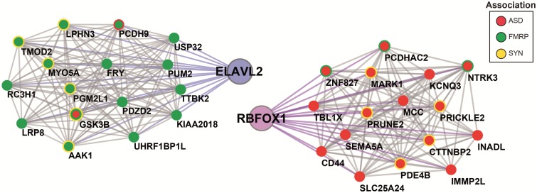
The role of post-transcriptional gene regulation in human brain development and neurodevelopmental disorders remains mostly uncharacterized. ELAV-like RNA-binding proteins (RNAbps) are a family of proteins that regulate several aspects of neuronal function including neuronal excitability and synaptic transmission, both critical to the normal function of the brain in cognition and behavior. Here, we identify the downstream neuronal transcriptional and splicing networks of ELAVL2, an RNAbp with previously unknown function in the brain. Expression of ELAVL2 was reduced in human neurons and RNA-sequencing was utilized to identify networks of differentially expressed and alternatively spliced genes resulting from haploinsufficient levels of ELAVL2. These networks contain a number of autism-relevant genes as well as previously identified targets of other important RNAbps implicated in autism spectrum disorder (ASD) including RBFOX1 and FMRP. ELAVL2-regulated co-expression networks are also enriched for neurodevelopmental and synaptic genes, and include genes with human-specific patterns of expression in the frontal pole. Together, these data suggest that ELAVL2 regulation of transcript expression is critical for neuronal function and clinically relevant to ASD.


Figures

Figure 1.
Figure 1.
ELAVL2 expression in human brain and phNs. (A) Expression distribution of RBFOX1 and ELAVL2 in fetal cortical regions (A1C, primary auditory cortex; DFC, dorsolateral frontal cortex; IPC, inferior parietal cortex; ITC, inferior temporal cortex; M1C, primary motor cortex; MFC, medial frontal cortex; OFC, orbital frontal cortex; S1C, primary sensory cortex; STC, superior temporal cortex; V1C, primary visual cortex; VFC, ventral frontal cortex). (B)ELAVL2 and RBFOX1 expression comparison in dorsolateral prefrontal cortex throughout the lifespan. The fetal developmental period in which RBFOX1 and ELAVL2 are strongly co-expressed is indicated. (C) Expression distribution of ELAVL2 and RBFOX1 in cortical layers (VZ, ventricular zone; SZ, subventricular zone; IZ, intermediate zone; SP, subplate; CP, cortical plate; MZ, marginal zone; SG, subpial granular layer). (D) Co-expression sub-module of RNAbps in prefrontal fetal cortex. Interactions represent potential protein interactions between RNAbps predicted by co-expressed genes. Each link corresponds to a weight calculated according to the correlation of the gene sets between nodes (see Methods section). RNAbps are preselected as nodes in the network. Shown are links between ELAVL2 (blue), RBFOX1 (purple), FMR1 (green) and other RNAbps (grey). (E) Co-localization confocal images of ELAVL2 and FMRP in phNs. Arrows indicate the perinuclear localization of both RNAbps. TO-PRO-3 is used to identify nuclei. Scale bar, 5 μm.
Figure 2.
Figure 2.
ELAVL2 kd alters alternative splicing events in phNs. (A) Characterization of ELAVL2 kd by immunoblotting. Protein expression of ELAVL2 was significantly reduced by 71% using shELAVL2. Samples were normalized to beta-actin and compared with shGFP control (t-test, P = 0.0012, n = 4 with each experiment performed at least in duplicate). (B) Protein–protein interaction network of ELAVL2 alternative spliced genes (blue) and RBFOX1 alternative spliced genes (purple). Also shown are genes associated with ASD (red); genes associated with FMRP-regulated pathways (green); and genes associated with synaptic function (yellow). (C) Overlaps of ELAVL2 and RBFOX1 alternatively spliced genes with ASD, FMRP-regulated pathways and synaptic genes. (D) Position enrichment of the ELAVL2 (blue) and RBFOX1- (purple) binding sites in exon-flanking intronic regions and the 3′UTR of alternative spliced genes for all of the alternatively spliced genes in either dataset. (E) Co-occurrence of binding sites in introns or UTRs of the 14 common alternatively spliced targets of ELAVL2 and RBFOX1. Red corresponds to a co-occurrence; Grey corresponds to a non-occurrence.
Figure 3.
Figure 3.
ELAVL2 kd alters gene expression in phNs. (A) Validation of 12 DEGs detected by RNA-seq using qRT-PCR. RNA-seq values are shown in blue and qPCR values are shown in grey. Standard error of the mean is indicated by the black bars. (B) Gene ontology enrichment of DEGs downstream of ELAVL2. (C)ELAVL2 DEGs are enriched for ASD genes. (D) Expression of the 13 ASD genes that are upregulated with ELAVL2 kd.
Figure 4.
Figure 4.
ELAVL2 kd alters genetic modules involved in neurodevelopmental networks. (A) ELAVL2 kd yielded two key modules (hEL13 and hEL24) enriched in synaptic genes, FMRP targets, and DEGs. Genes with higher connectivity are indicated by larger node size. ASD genes (red); FMRP targets (green); synaptic genes (yellow); ELAVL2 DEGs (blue); RBFOX1 DEGs (purple); Human frontal pole-specific genes derived by comparison of gene expression in human and non-human primate brains (black) (25). Barplots below each module show the relationship of each sample to the module eigengene. In both modules, ELAVL2 kd samples show strong positive correlation. (B) Gene ontology enrichment indicates modules strongly enriched for neurogenesis and neurodevelopmental pathways. (C) Preservation of the ELAVL2 modules in the RBFOX1 kd data set (significance Z score > 5). The two key modules of interest are indicated.
Figure 5.
Figure 5.
ELAVL2 kd alters co-expression of genes involved in synaptic, ASD, and FMRP pathways. On the left, the co-expressed genes with ELAVL2; on the right, the co-expressed genes with RBFOX1. ASD genes (red); FMRP targets (green); Synaptic genes (yellow).

References

    1. De Rubeis S., He X., Goldberg A.P., Poultney C.S., Samocha K., Cicek A.E., Kou Y., Liu L., Fromer M., Walker S. et al. (2014) Synaptic, transcriptional and chromatin genes disrupted in autism. Nature, 515, 209–215. - PMC - PubMed
    1. Butler M.G., Rafi S.K., Hossain W., Stephan D.A., Manzardo A.M. (2015) Whole exome sequencing in females with autism implicates novel and candidate genes. Int. J. Mol. Sci., 16, 1312–1335. - PMC - PubMed
    1. Sanders S.J., Murtha M.T., Gupta A.R., Murdoch J.D., Raubeson M.J., Willsey A.J., Ercan-Sencicek A.G., DiLullo N.M., Parikshak N.N., Stein J.L. et al. (2012) De novo mutations revealed by whole-exome sequencing are strongly associated with autism. Nature, 485, 237–241. - PMC - PubMed
    1. Yu T.W., Chahrour M.H., Coulter M.E., Jiralerspong S., Okamura-Ikeda K., Ataman B., Schmitz-Abe K., Harmin D.A., Adli M., Malik A.N. et al. (2013) Using whole-exome sequencing to identify inherited causes of autism. Neuron, 77, 259–273. - PMC - PubMed
    1. Raj B., Blencowe B.J. (2015) Alternative splicing in the mammalian nervous system: recent insights into mechanisms and functional roles. Neuron, 87, 14–27. - PubMed

Publication types

MeSH terms