Review
. 2016 Nov:55:52-81.
doi: 10.1016/j.preteyeres.2016.05.003. Epub 2016 Jun 1.

Molecular basis for photoreceptor outer segment architecture

Affiliations
Review

Molecular basis for photoreceptor outer segment architecture

Andrew F X Goldberg et al. Prog Retin Eye Res. 2016 Nov.

Abstract

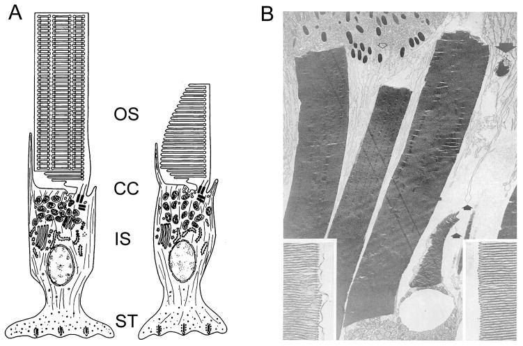
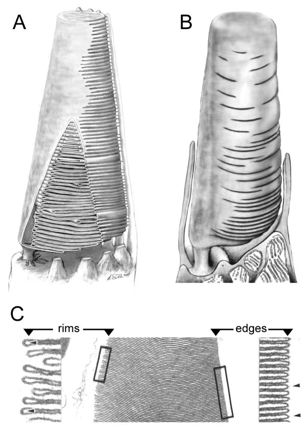
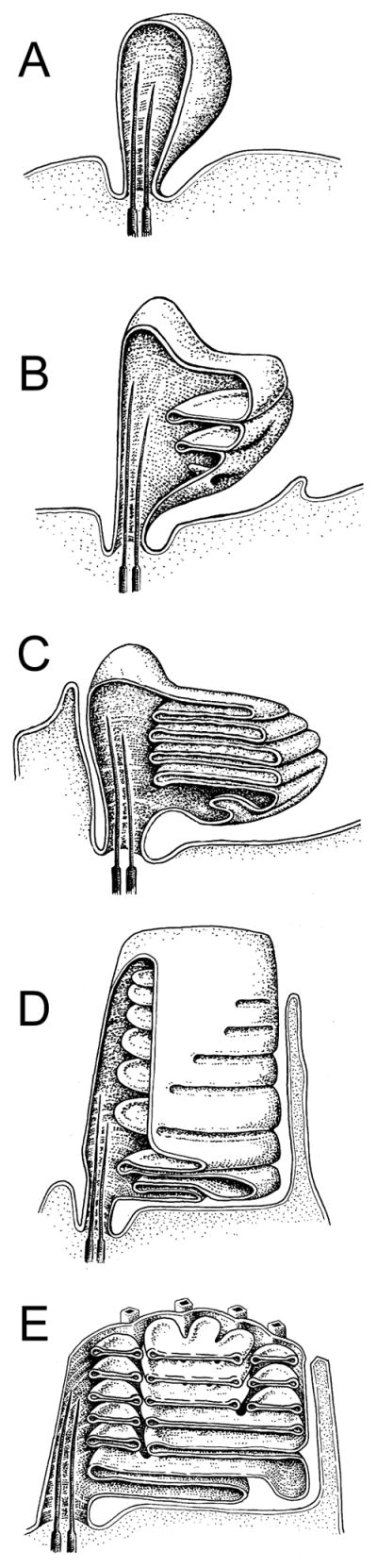
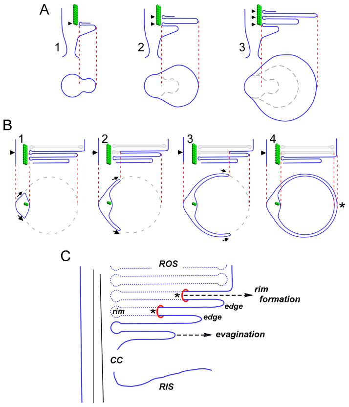
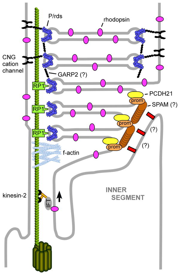
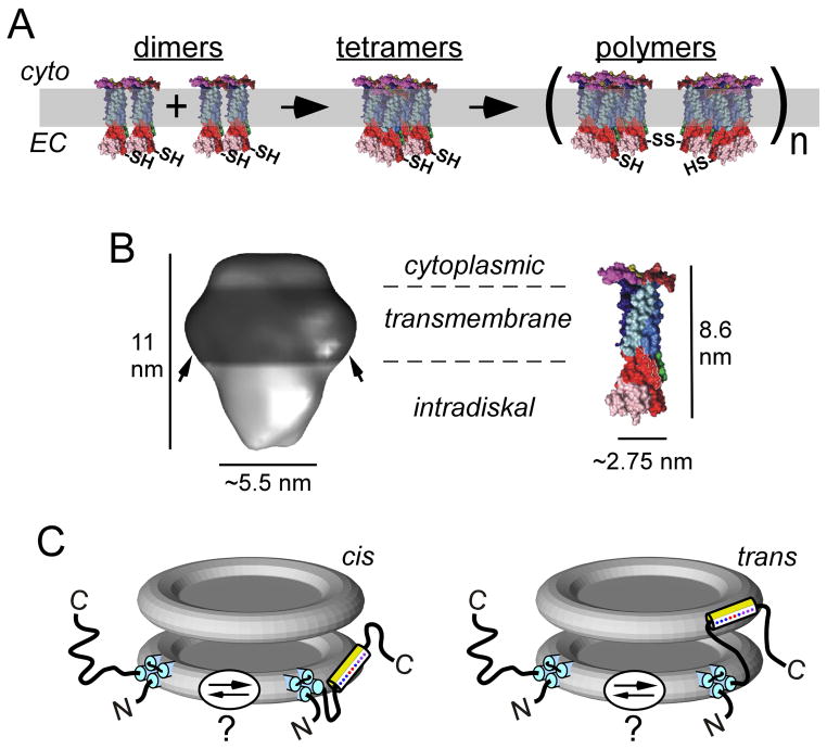
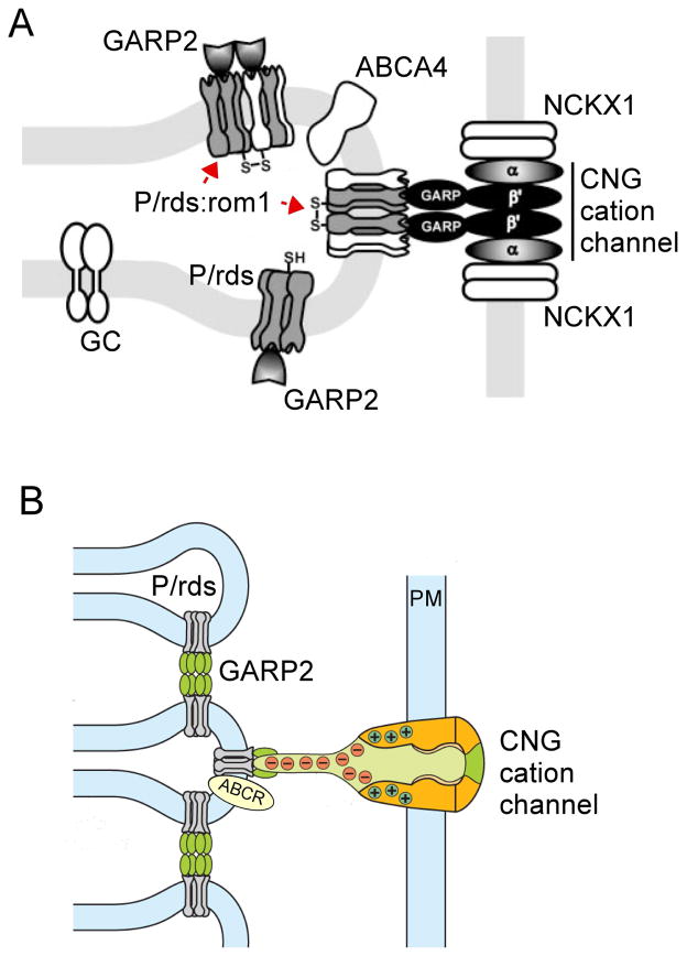
To serve vision, vertebrate rod and cone photoreceptors must detect photons, convert the light stimuli into cellular signals, and then convey the encoded information to downstream neurons. Rods and cones are sensory neurons that each rely on specialized ciliary organelles to detect light. These organelles, called outer segments, possess elaborate architectures that include many hundreds of light-sensitive membranous disks arrayed one atop another in precise register. These stacked disks capture light and initiate the chain of molecular and cellular events that underlie normal vision. Outer segment organization is challenged by an inherently dynamic nature; these organelles are subject to a renewal process that replaces a significant fraction of their disks (up to ∼10%) on a daily basis. In addition, a broad range of environmental and genetic insults can disrupt outer segment morphology to impair photoreceptor function and viability. In this chapter, we survey the major progress that has been made for understanding the molecular basis of outer segment architecture. We also discuss key aspects of organelle lipid and protein composition, and highlight distributions, interactions, and potential structural functions of key OS-resident molecules, including: kinesin-2, actin, RP1, prominin-1, protocadherin 21, peripherin-2/rds, rom-1, glutamic acid-rich proteins, and rhodopsin. Finally, we identify key knowledge gaps and challenges that remain for understanding how normal outer segment architecture is established and maintained.

Keywords: Cilia; Membrane curvature; Outer segment; Photoreceptor; Retinal degeneration; Tetraspanin.


Figures

Figure 1
Figure 1. The OS in context
A) Illustrations of rod (left) and cone (right) frog photoreceptors; adapted from (Bok, 1985). In each subtype, the light-sensitive OS is attached to the inner segment (IS) via the connecting cilium (CC). B) Transmission electron microscopy of adult frog OSs; three rod and one cone OSs are present. Apical processes (small arrows) and a phagosome containing shed disks (large arrow) are present. Insets show enlarged images of disk rims (left) and edges (right) from a cone OS. Reproduced with permission from (Kinney and Fisher, 1978a).
Figure 2
Figure 2. OS disk membrane topology varies by cell subtype, species, and disk age
Renderings of cone photoreceptor OSs (A, B; not to scale). A) frog cone adapted from (Weber et al., 2011). Frog cone OSs possess a strong basal-to-distal taper. A portion of the plasma membrane has been cut away to reveal the internal structure. The plasma membrane opposite the eccentrically positioned cilium is pleated to form a large stack of disks that are further defined by the presence of rims, which partially bound the pleats and begin the process of defining a new compartment. The apertures that expose disk surfaces to the extracellular milieu extend about halfway around the disk boundaries, and are aligned along the length of the OS to those of adjacent disks. B) Mammalian cone adapted from (Anderson et al., 1978). Mammalian cone OSs possess more subtle basal-to-distal tapers than those present in frog cones. Like frog cone OSs, mammalian cone OSs also possess disks that remain in continuity with the plasma membrane. In this case however, the boundary of each mature disk is largely differentiated from the plasma membrane as a rim, so that the disks are largely internalized and only relatively small apertures remain open to the extracellular milieu. Moreover, these apertures are not aligned along the OS length, and make topological interpretation of sectioned tissue quite challenging. C) Transverse sections of frog cone OSs illustrate the distinction between U-shaped disk edges (right) and hairpin-shaped disk rims (left) that are present in the partially-internalized disks prevalent in cones. Adapted from (Corless and Fetter, 1987).
Figure 3
Figure 3. Development of the vertebrate OS
Renderings of ROS morphogenesis in Rana pipians tadpole; adapted from (Nilsson, 1964). Retinal pigment epithelium is omitted for clarity. A) Ballooning of the ciliary plasma membrane. B) Continued plasma membrane expansion and invagination forms initial nascent disk membranes. C) Expansion and displacement of existing disks by new disks create plasma membrane pleating and an initial stack of open disks. D) Disks are further differentiated by the gradual expansion of rims, which begin the process of internalization. E) Development of incisures in mature, completely internalized disks. Cone OS development was proposed to be similar, but rim expansion halts prior to complete disk internalization, and no incisures are formed.
Figure 4
Figure 4. Steinberg et al. two-step (A, B) model for OS disk morphogenesis
A) Step1 - growth of the ciliary plasma membrane creates an evagination with an upper surface anchored at an axonemal microtubule (A.1). Anchoring of the evagination lower surface allows for initiation of a new evagination (A.2). Evagination translation towards the cilium distal tip is accompanied by flattening and growth to mature disk diameter (A.3). At this stage, disks are bounded by edges; rims are absent, except for those regions immediately adjacent to the axonemal microtubules (arrowheads). Below: Tangential views sectioned through of each new evagination (arrowheads in longitudinal views). B) Step 2 - disk internalization begins by expansion of the rim region that anchors evaginations to the axoneme. Above: longitudinal views show expansion of the disk rim from left to right across one open disk (arrowheads). Below: tangential views sectioned through a single disk (arrowheads) undergoing internalization. The disk rim present at the axoneme advances bilaterally, in parallel with the enclosing plasma membrane at two growth points (arrows). Disk rim growth is proposed to occur by membrane addition. Completion of rim formation and disk internalization occurs opposite the axoneme (asterisk), and requires a membrane fission event to sever disk-plasma membrane continuity. Adapted from (Steinberg et al., 1980). C) A view of disk morphogenesis that emphasizes disk rim formation via membrane addition. Asterisks illustrate growth points, at which the addition of new plasma membrane allows rim formation to advance. Adapted from (Arikawa et al., 1992).
Figure 5
Figure 5. Tethering features that may contribute to OS architecture
A) TEM image of a frog cone OS prepared by freeze-fracture deep-etch rotary-shadowing. Numerous regularly-organized axially-oriented tethers link adjacent disk edges. The leading edge of the OS plasma membrane (PM), the point at which disk internalization has halted, is indicated between the arrows (upper left). Several calycal processes (CP) are present in the image. Image adapted from (Fetter and Corless, 1987). B) Fracturing and removal of plasma membrane (PM) from a similarly prepared toad rod OS exposes regularly organized tethers linking adjacent disk rims. Image adapted from (Roof and Heuser, 1982). C) A similar preparation technique applied to a bovine rod OS protein reveals tethers between disk rims and the plasma membrane (PM). Image adapted from (Roof and Heuser, 1982). D) Cryoelectron microscopy of frozen vitrified mouse rod OSs finds tethers (orange) between disk rims and the plasma membrane (PM; blue) and “spacers” (red; arrow) distributed randomly upon the lamellar portion of the disk. Image adapted from (Nickell et al., 2007).
Figure 6
Figure 6. Organization of nascent rod OS disk membranes
A) Evaginating basal disks of mouse rod OSs (arrows) show clear continuity with the ciliary plasma membrane when retinas are preserved via transcardial perfusion with fixative prior to dissection; adapted from (Volland et al., 2015). Arrows identify nascent disks continuous with the plasma. B) 3D rendering of an electron tomogram of the basal region of a monkey rod OS. Nascent disks, which show continuity with the ciliary plasma membrane are colored green, while fully internalized mature disks are colored blue; adapted from (Volland et al., 2015). C–E) In vivo perfusion with tannic acid-containing fixative highlights open disks; adapted from (Ding et al., 2015). The boxed region in C) is enlarged in D) and illustrated as a pseudo-colored tracing in E). Arrowheads mark nascent disks; arrows identify mature disks. The poor permeability of tannic acid through membranes results in enhanced staining of disks that retain continuity with the plasma membrane. F–G) Linkages connect evagination (nascent disk) edges with IS plasma membrane; adapted from (Burgoyne et al., 2015). F) Visualization of an OS base from a single tomogram slice. A 3D model of the tomographic data from the boxed area in F) is presented in G). The model illustrates fibers (red) linking an evagination (green) edge to the IS plasma membrane (yellow).
Figure 7
Figure 7. General mechanisms for the generation of membrane curvature
Cells actively control membrane curvature using a variety of mechanisms. Although membrane lipid (A, B) and protein (C–F) composition can each contribute to curvature generation, the bulk of the energy required for shaping high curvature membranes is provided by specialized curvature-generating proteins. Mechanisms that contribute to membrane curvature generation include, A) phospholipid headgroup composition, B) phospholipid acyl chain composition, C) protein scaffolding, D) amphipathic helix insertion, E) hydrophobic loop insertion, F) conically-shaped transmembrane proteins.
Figure 8
Figure 8. Distributions of OS-resident proteins with likely roles for organelle structure
Schematic drawing (not to scale) of the basal rod OS shows the locations of the proteins discussed in this article. Kinesin-2 (black/gold) drives anterograde transport of IFT particles and cargo into the OS. Retrograde IFT transport (by dynein) is not illustrated. F-actin (light blue) is localized at the OS base in association with axonemal microtubules. These microfilaments are required for the initiation of new disk evaginations. Rhodopsin (magenta) is present in the ciliary plasma membrane, OS plasma membrane, and OS disk lamellar membranes. Rhodopsin abundance has a dose-dependent effect on disk diameter. Prominin-1 (prom; orange) is present at the edges of basal evaginations, where it interacts with PCDH21 and potentially SPAM. Prominin-1 may function to maintain disk edge membrane curvature. SPAM (brown) localization is putative, and based on its documented interaction with prominin-1 in Drosophila photoreceptors. SPAM function remains to be determined. PCDH21 (yellow) is present at the edges of basal evaginations, where it interacts with prominin-1. The identity (and interactions) of tethers (red rectangles), which link nascent disk edges and the IS plasma membrane remains to be determined. P/rds and P/rds-rom1 complexes (dark blue) are present in disk rim domains. P/rds likely plays a direct role for generating membrane curvature and may participate in disk-disk tethering. Rom-1 modulates P/rds function. RP1 (light green) is associated with axonemal microtubules at the IS-OS junction. RP1 may link nascent OS disks to the axoneme to govern their morphogenesis and stacking. The CNG cation channel (black) is present in the OS plasma membrane. In addition to its primary role for regulating OS permeability, the CNG cation channel contributes to OS structural stability by tethering the plasma membrane to disk rims via interaction with P/rds. GARP-2 (black) is present at OS disk rims. It may contribute to the long-term structural stability of OS disk stacking interactions and/or cGMP phosphodiesterase localization (not shown).
Figure 9
Figure 9. P/rds oligomerization, gross structure, and topology at rod OS disk rims
A) P/rds dimers (modeled using CD81 monomers, described below) are likely assembled at the biosynthetic level and must undergo additional steps of oligomerization to function in support of OS morphogenesis. Adapted from (Goldberg, 2006). B) Left: 3D structure of the detergent-solubilized P/rds:rom1 heterotetramer. Arrows indicate a presumed annulus of detergent molecules bound at the transmembrane domains. Adapted from (Kevany et al., 2013). Right: 3D model of CD81, a homologous tetraspanin with a mass (26 kDa) somewhat smaller than that of P/rds (39 kDa). Image adapted from (Seigneuret, 2006). The dimensions determined for the P/rds:rom1 heterotetramer are consistent with a complex of four tetraspanin monomers. C) Topology of P/rds in the disk rim, showing hypothesized partitioning of the C-terminal amphipathic helix. The amphipathic helix is leashed to the P/rds transmembrane domain by a disordered region sufficient to bridge adjacent disks, potentially allowing trans insertion.
Figure 10
Figure 10. Disk rim complexes proposed to contribute to OS organization
Models proposed for disk rim scaffolding of OS components, based on localization and protein-protein interaction data. A) Interactions between GARPs and P/rds are proposed to mediate fibril linkages between disk rims and the OS plasma membrane. Adapted from (Poetsch et al., 2001). B) An elaboration of the Poetsch et al. model suggests GARP2 interaction with P/rds mediates disk-disk stacking. Adapted from (Kaupp and Seifert, 2002).

References

    1. Abd El-Aziz MM, Barragan I, O’Driscoll CA, Goodstadt L, Prigmore E, Borrego S, Mena M, Pieras JI, El-Ashry MF, Safieh LA, Shah A, Cheetham ME, Carter NP, Chakarova C, Ponting CP, Bhattacharya SS, Antinolo G. EYS, encoding an ortholog of Drosophila spacemaker, is mutated in autosomal recessive retinitis pigmentosa. Nat Genet. 2008;40:1285–1287. - PMC - PubMed
    1. Agbaga MM, DK, Brush RS, Lydic T, Conley SM, Naash MI, Gavin RE, Busik JV, Anderson RE. Differential composition of docosahexaenoic acid and very long chain polyunsaturated fatty acids in rod and cone photoreceptor membranes. Invest Ophthalmol Vis Sci. 2014;55:370.
    1. Albert AD, Boesze-Battaglia K. The role of cholesterol in rod outer segment membranes. Prog Lipid Res. 2005;44:99–124. - PMC - PubMed
    1. Anderson DH, Fisher SK, Steinberg RH. Mammalian cones: disc shedding, phagocytosis, and renewal. Invest Ophthalmol Vis Sci. 1978;17:117–133. - PubMed
    1. Anderson RE, Maude MB. Phospholipids of bovine outer segments. Biochem. 1970;9:3624–3628. - PubMed

Publication types