. 2017 Feb;39(1):56-65.
doi: 10.1111/ics.12348. Epub 2016 Jul 4.

Molecular basis of retinol anti-ageing properties in naturally aged human skin in vivo

Affiliations

Molecular basis of retinol anti-ageing properties in naturally aged human skin in vivo

Y Shao et al. Int J Cosmet Sci. 2017 Feb.

Abstract

Objective: Retinoic acid has been shown to improve the aged-appearing skin. However, less is known about the anti-ageing effects of retinol (ROL, vitamin A), a precursor of retinoic acid, in aged human skin in vivo. This study aimed to investigate the molecular basis of ROL anti-ageing properties in naturally aged human skin in vivo.

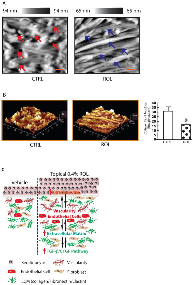
Methods: Sun-protected buttock skin (76 ± 6 years old, n = 12) was topically treated with 0.4% ROL and its vehicle for 7 days. The effects of topical ROL on skin epidermis and dermis were evaluated by immunohistochemistry, in situ hybridization, Northern analysis, real-time RT-PCR and Western analysis. Collagen fibrils nanoscale structure and surface topology were analysed by atomic force microscopy.

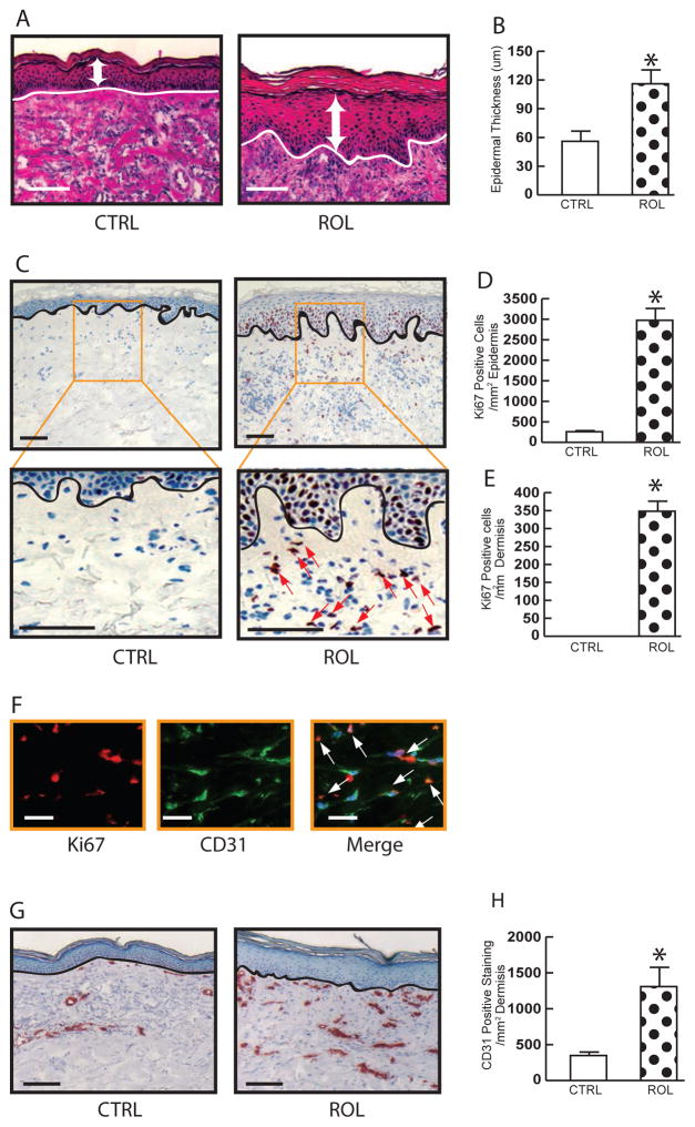
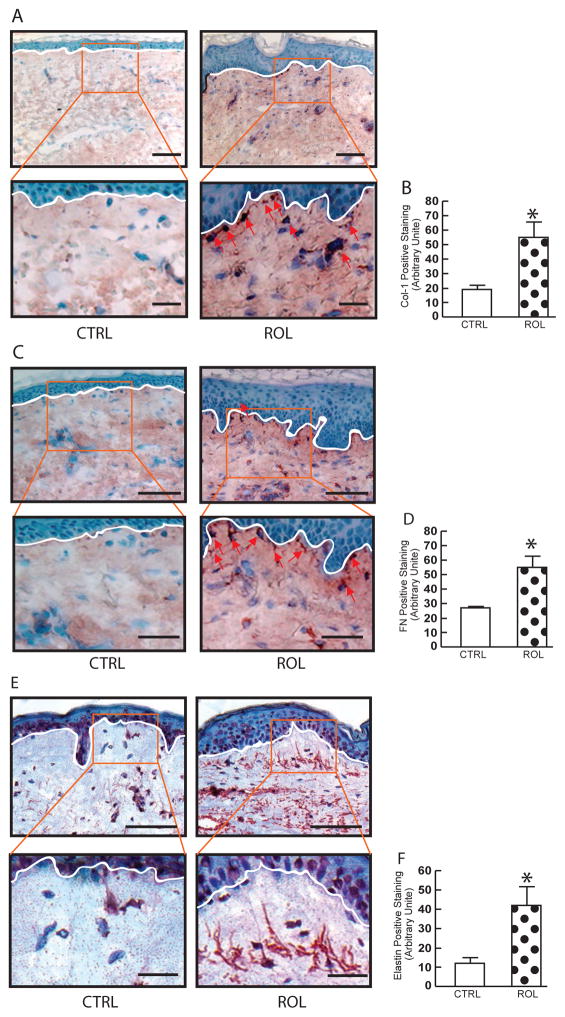
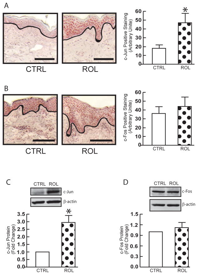
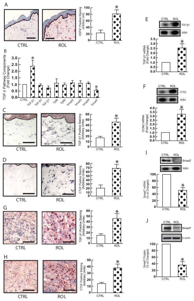
Results: Topical ROL shows remarkable anti-ageing effects through three major types of skin cells: epidermal keratinocytes, dermal endothelial cells and fibroblasts. Topical ROL significantly increased epidermal thickness by stimulating keratinocytes proliferation and upregulation of c-Jun transcription factor. In addition to epidermal changes, topical ROL significantly improved dermal extracellular matrix (ECM) microenvironment; increasing dermal vascularity by stimulating endothelial cells proliferation and ECM production (type I collagen, fibronectin and elastin) by activating dermal fibroblasts. Topical ROL also stimulates TGF-β/CTGF pathway, the major regulator of ECM homeostasis, and thus enriched the deposition of ECM in aged human skin in vivo. 0.4% topical ROL achieved similar results as seen with topical retinoic acid, the biologically active form of ROL, without causing noticeable signs of retinoid side effects.

Conclusion: 0.4% topical ROL shows remarkable anti-ageing effects through improvement of the homeostasis of epidermis and dermis by stimulating the proliferation of keratinocytes and endothelial cells, and activating dermal fibroblasts. These data provide evidence that 0.4% topical ROL is a promising and safe treatment to improve the naturally aged human skin.

Keywords: TGF-β; extracellular matrix; retinoid; skin ageing; vascularity.


Figures

Figure 1
Figure 1
Topical ROL increases epidermal thickness and dermal vascularity by proliferation of epidermal keratinocytes and dermal endothelial cells, respectively, in aged human skin in vivo. OCT-embedded skin sections (7μm) were obtained from aged (76±6 years) healthy sun-protected buttock skin after topical treatment of vehicle and 0.4% retinol for seven days. (A) H&E staining. Representative images of twelve individuals (N=12). White arrows indicate epidermal thickness. Bars=100μm. (B) Quantification of epidermal thickness (μm). (C) Ki67 immunostaining. 3.0× enlargement of the boxed region is shown to lower panels. Bars= 100μm. Representative images of twelve individuals (N=12). Quantification of epidermal (D) and dermal (E) Ki 67 immunostaining. (F) Ki67 and CD31 co-immunofluoresce staining. Skin sections were co-immunofluoresce stained with Ki67 and CD31, a marker of endothelial cells. Arrows indicate double stained cells. Representative of five individuals. Bar=50μm. (G) CD31 immunostaining. Representative images of twelve individuals (N=12). Bar=100μm. (H) Quantification of CD31 immunostaining. All immunostainings were quantified by computerized image analysis (Image-pro Plus software, version 4.1, Media Cybernetics, MD) and data are expressed as mean±SEM, *p<0.05. N=12.
Figure 2
Figure 2
ROL improves dermal ECM microenvironment in aged human skin in vivo. OCT-embedded skin sections (7μm) were obtained from aged (76±6 years) healthy sun-protected buttock skin after topical treatment of vehicle and 0.4% retinol for seven days. (A) Type I procollagen immunostaining. 3× enlargement of the boxed region is shown to lower panels. Representative images of twelve individuals (N=12). Arrows indicate positive cells. Bars=100μm. (B) Quantification of type I procollagen. (C) Fibronectin immunostaining. 2.5× enlargement of the boxed region is shown to lower panels. Representative images of twelve individuals (N=12). Arrows indicate positive cells. Bars= 100μm. (D) Quantification of fibronectin. (E) Tropoelastin immunostaining. 3.0× enlargement of the boxed region is shown to lower panels. Representative images of twelve individuals (N=12). Bars=100μm. (F) Quantification of tropoelastin. All immunostainings were quantified by computerized image analysis (Image-pro Plus software, version 4.1, Media Cybernetics, MD) and data are expressed as mean±SEM, *p<0.05. N=12.
Figure 3
Figure 3
Elevated epidermal-specific c-Jun transcription factor by topical ROL in aged human skin in vivo. OCT-embedded skin sections (7μm) were obtained from aged (76±6 years) healthy sun-protected buttock skin after topical treatment of vehicle and 0.4% retinol for seven days. (A) c-Jun immunostaining. Representative images of twelve individuals (N=12). Bars=100μm. (B) c-Fos immunostaining. Representative images of twelve individuals (N=12). Bars=100μm. All immunostainings were quantified by computerized image analysis (Image-pro Plus software, version 4.1, Media Cybernetics, MD) and data are expressed as mean±SEM, *p<0.05. N=12. (C) c-Jun protein. (D) c-Fos protein. Protein levels were determined by Western analysis. Protein levels were normalized to β-actin (loading control). Insets show representative Western blots. Mean ± SEM, N=6, *p < 0.05.
Figure 4
Figure 4
Topical ROL stimulates TGF-β/CTGF pathway, the major regulator of ECM homeostasis, in aged human skin in vivo. OCT-embedded skin sections (7μm) were obtained from aged (76±6 years) healthy sun-protected buttock skin after topical treatment of vehicle and 0.4% retinol for seven days. (A) HSP47 immunostaining. Representative images of twelve individuals (N=12). Bars=100μm. (B) TGF-β pathway components mRNA levels. Total RNA was prepared from human skin samples. mRNA levels were determined by real-time RT-PCR. mRNA levels were normalized to 36B4 (internal housekeeping gene control). Mean ± SEM, N=12, *p<0.05. (C) TGF-β1 (D) CTGF/CCN2 in situ hybridization. Representative images of twelve individuals (N=12). Bars= 100μm. (E) TGF-β1 (F) CTGF/CCN2 Northern analysis. The intensities were quantified and normalized using 36B4 as loading control. Insets show representative Northern blots. Data are expressed as mean±SEM, *p<0.05. N=12. (G) TGF-β1 (H) CTGF/CCN2 immunostaining. Representative images of twelve individuals (N=12). Bars= 100μm. (I) Smad7 Northern analysis. The intensities were quantified and normalized using 36B4 as loading control. Inset shows representative Northern blots. Data are expressed as mean±SEM, *p<0.05. N=12. (J) Smad7 protein. Smad7 protein levels were determined by Western analysis. Protein levels were normalized to β-actin (loading control). Insets show representative Western blots. Mean ± SEM, N =6, *p < 0.05. All positive staining was quantified by computerized image analysis (Image-pro Plus software, version 4.1, Media Cybernetics, MD). Data are expressed as mean±SEM, *p<0.05. N=12.
Figure 5
Figure 5
Deposition of mature collagen is increased by topical ROL in aged human skin in vivo. OCT-embedded skin sections (7μm) were obtained from aged (76±6 years) healthy sun-protected buttock skin after topical treatment of vehicle and 0.4% retinol for seven days. (A) Nanoscale collagen fibrils were imaged by AFM. Representative AFM images were shown. N=12. The blue arrows indicate intact collagen fibrils and red arrows heads indicate damaged collagen fibrils. (B) Three dimensional collagen fibrils. Dermal roughness was analyzed using Nanoscope Analysis software (Nanoscope_Analysis_v120R1sr3, Bruker-AXS, Santa Barbara, CA). All results are expressed as the mean ± SEM, N=12, *p < 0.05. Bars=100 nm. (C) Proposed model for topical ROL exerts anti-aging effects in aged human skin by proliferation of epidermal keratinocytes and dermal endothelial cells and activation of dermal fibroblasts (see Discussion for details).

References

    1. Quan T. Skin connective tissue aging and dermal fibroblasts. Dermal Fibroblasts: Histological Perspectives, Characterization and Role in Disease. 2013:31–55.
    1. Fisher GJ, et al. Mechanisms of photoaging and chronological skin aging. Arch Dermatol. 2002;138(11):1462–70. - PubMed
    1. Thomas DR, Burkemper NM. Aging skin and wound healing. Clin Geriatr Med. 2013;29(2):xi–xx. - PubMed
    1. Worley CA. Aging skin and wound healing. Dermatol Nurs. 2006;18(3):265–6. - PubMed
    1. Bissell MJ, Hines WC. Why don’t we get more cancer? A proposed role of the microenvironment in restraining cancer progression. Nat Med. 2011;17(3):320–9. - PMC - PubMed