

Remodeling the Vascular Microenvironment of Glioblastoma with α-Particles
- PMID: 27261519
- PMCID: PMC5093034
- DOI: 10.2967/jnumed.116.173559
Remodeling the Vascular Microenvironment of Glioblastoma with α-Particles
Abstract
Tumors escape antiangiogenic therapy by activation of proangiogenic signaling pathways. Bevacizumab is approved for the treatment of recurrent glioblastoma, but patients inevitably develop resistance to this angiogenic inhibitor. We previously investigated targeted α-particle therapy with 225Ac-E4G10 as an antivascular approach and showed increased survival and tumor control in a high-grade transgenic orthotopic glioblastoma model. Here, we investigated changes in tumor vascular morphology and functionality caused by 225Ac-E4G10.
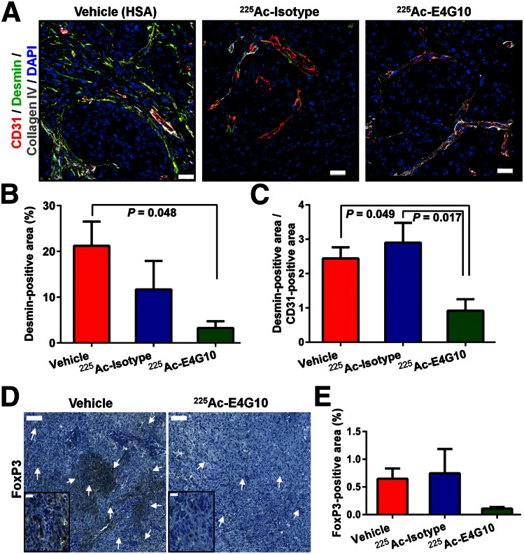
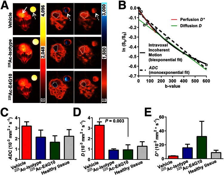
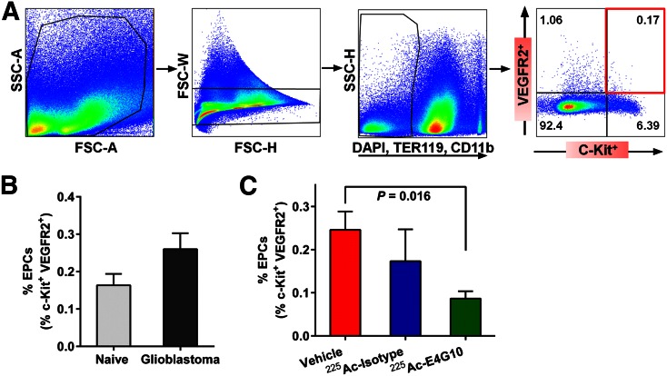
Methods: We investigated remodeling of the tumor microenvironment in transgenic Ntva glioblastoma mice using a therapeutic 7.4-kBq dose of 225Ac-E4G10. Immunofluorescence and immunohistochemical analyses imaged morphologic changes in the tumor blood-brain barrier microenvironment. Multicolor flow cytometry quantified the endothelial progenitor cell population in the bone marrow. Diffusion-weighted MR imaged functional changes in the tumor vascular network.
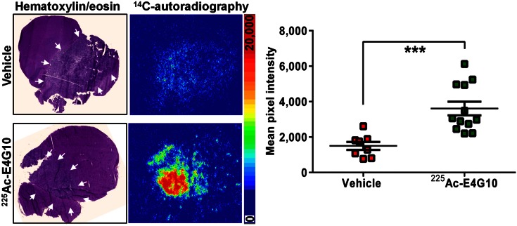
Results: The mechanism of drug action is a combination of remodeling of the glioblastoma vascular microenvironment, relief of edema, and depletion of regulatory T and endothelial progenitor cells. The primary remodeling event is the reduction of both endothelial and perivascular cell populations. Tumor-associated edema and necrosis were lessened, resulting in increased perfusion and reduced diffusion. Pharmacologic uptake of dasatinib into tumor was enhanced after α-particle therapy.
Conclusion: Targeted antivascular α-particle radiation remodels the glioblastoma vascular microenvironment via a multimodal mechanism of action and provides insight into the vascular architecture of platelet-derived growth factor-driven glioblastoma.
Keywords: 225Ac; actinium-225; glioblastoma; pericytes; radioimmunotherapy; vascular endothelium.
© 2016 by the Society of Nuclear Medicine and Molecular Imaging, Inc.
Conflict of interest statement
of Potential Conflicts of Interest M. R. McDevitt and D. A. Scheinberg declare associations with Actinium Pharmaceuticals, Inc.
Figures
Similar articles
-
Vascular Targeted Radioimmunotherapy for the Treatment of Glioblastoma.J Nucl Med. 2016 Oct;57(10):1576-1582. doi: 10.2967/jnumed.115.171371. Epub 2016 Apr 28. J Nucl Med. 2016. PMID: 27127217 Free PMC article.
-
Selective alpha-particle mediated depletion of tumor vasculature with vascular normalization.PLoS One. 2007 Mar 7;2(3):e267. doi: 10.1371/journal.pone.0000267. PLoS One. 2007. PMID: 17342201 Free PMC article.
-
IL13RA2 targeted alpha particle therapy against glioblastomas.Oncotarget. 2017 Jun 27;8(26):42997-43007. doi: 10.18632/oncotarget.17792. Oncotarget. 2017. PMID: 28562337 Free PMC article.
-
[The stem cell niche in glioblastoma: from fundamental aspects to targeted therapies].Bull Cancer. 2015 Jan;102(1):24-33. doi: 10.1016/j.bulcan.2014.07.001. Epub 2015 Jan 6. Bull Cancer. 2015. PMID: 25609493 Review. French.
-
The glioblastoma vasculature as a target for cancer therapy.Biochem Soc Trans. 2014 Dec;42(6):1647-52. doi: 10.1042/BST20140278. Biochem Soc Trans. 2014. PMID: 25399584 Review.
Cited by
-
Leveraging Bioorthogonal Click Chemistry to Improve 225Ac-Radioimmunotherapy of Pancreatic Ductal Adenocarcinoma.Clin Cancer Res. 2019 Jan 15;25(2):868-880. doi: 10.1158/1078-0432.CCR-18-1650. Epub 2018 Oct 23. Clin Cancer Res. 2019. PMID: 30352909 Free PMC article.
-
Beyond the Barrier: Targeted Radionuclide Therapy in Brain Tumors and Metastases.Pharmaceutics. 2019 Aug 1;11(8):376. doi: 10.3390/pharmaceutics11080376. Pharmaceutics. 2019. PMID: 31374991 Free PMC article. Review.
-
Brain intratumoural astatine-211 radiotherapy targeting syndecan-1 leads to durable glioblastoma remission and immune memory in female mice.EBioMedicine. 2024 Jul;105:105202. doi: 10.1016/j.ebiom.2024.105202. Epub 2024 Jun 20. EBioMedicine. 2024. PMID: 38905749 Free PMC article.
-
Current landscape and future directions of targeted-alpha-therapy for glioblastoma treatment.Theranostics. 2025 Mar 31;15(11):4861-4889. doi: 10.7150/thno.106081. eCollection 2025. Theranostics. 2025. PMID: 40303349 Free PMC article. Review.
-
Potential for Nuclear Medicine Therapy for Glioblastoma Treatment.Front Pharmacol. 2019 Jul 10;10:772. doi: 10.3389/fphar.2019.00772. eCollection 2019. Front Pharmacol. 2019. PMID: 31354487 Free PMC article. Review.
References
MeSH terms
Substances
Grants and funding
LinkOut - more resources
Full Text Sources
Other Literature Sources
Medical