

Four-dimensional MRI flow examinations in cerebral and extracerebral vessels - ready for clinical routine?
- PMID: 27262148
- PMCID: PMC4939804
- DOI: 10.1097/WCO.0000000000000341
Four-dimensional MRI flow examinations in cerebral and extracerebral vessels - ready for clinical routine?
Abstract
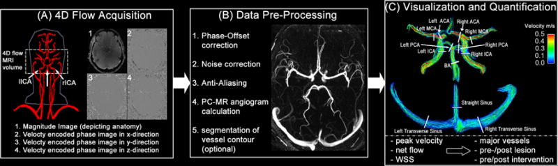
Purpose of review: To evaluate the feasibility of 4-dimensional (4D) flow MRI for the clinical assessment of cerebral and extracerebral vascular hemodynamics in patients with neurovascular disease.
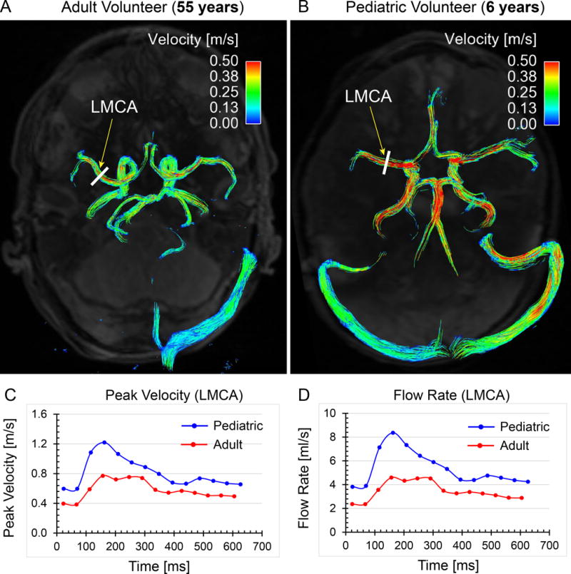
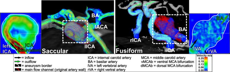
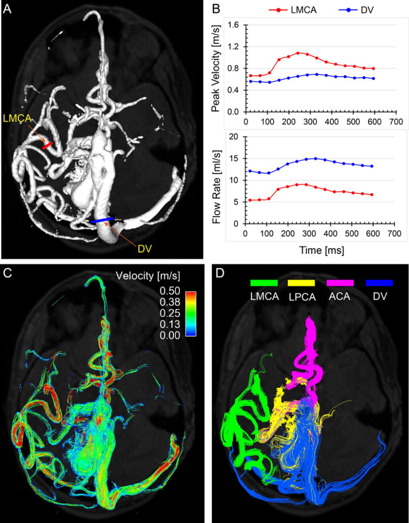
Recent findings: 4D flow MRI has been applied in multiple studies to qualitatively and quantitatively study intracranial aneurysm blood flow for potential risk stratification and to assess treatment efficacy of various neurovascular lesions, including intraaneurysmal and parent artery blood flow after flow diverter stent placement and staged embolizations of arteriovenous malformations and vein of Galen aneurysmal malformations. Recently, the technique has been utilized to characterize age-related changes of normal cerebral hemodynamics in healthy individuals over a broad age range.
Summary: 4D flow MRI is a useful tool for the noninvasive, volumetric and quantitative hemodynamic assessment of neurovascular disease without the need for gadolinium contrast agents. Further improvements are warranted to overcome technical limitations before broader clinical implementation. Current developments, such as advanced acceleration techniques (parallel imaging and compressed sensing) for faster data acquisition, dual or multiple velocity encoding strategies for more accurate arterial and venous flow quantification, ultrahigh-field strengths to achieve higher spatial resolution and streamlined postprocessing workflow for more efficient and standardized flow analysis, are promising advancements in 4D flow MRI.
Conflict of interest statement
There are no conflicts of interest to report.
Figures
References
-
- Atlas SW. Magnetic resonance imaging of intracranial aneurysms. Neuroimaging clinics of North America. 1997;7(4):709–20. - PubMed
-
- Frangos SG, Gahtan V, Sumpio B. Localization of atherosclerosis: role of hemodynamics. Arch Surg. 1999;134(10):1142–9. - PubMed
-
- Prado CM, Ramos SG, Alves-Filho JC, Elias J, Jr, Cunha FQ, Rossi MA. Turbulent flow/low wall shear stress and stretch differentially affect aorta remodeling in rats. Journal of hypertension. 2006;24(3):503–15. - PubMed
-
- Zarins CK, Giddens DP, Bharadvaj BK, Sottiurai VS, Mabon RF, Glagov S. Carotid bifurcation atherosclerosis. Quantitative correlation of plaque localization with flow velocity profiles and wall shear stress. Circulation research. 1983;53(4):502–14. - PubMed
Publication types
MeSH terms
Grants and funding
LinkOut - more resources
Full Text Sources
Other Literature Sources
Medical
Research Materials
