

Tissue-Level Mechanical Properties of Bone Contributing to Fracture Risk
- PMID: 27263108
- PMCID: PMC4927361
- DOI: 10.1007/s11914-016-0314-3
Tissue-Level Mechanical Properties of Bone Contributing to Fracture Risk
Abstract
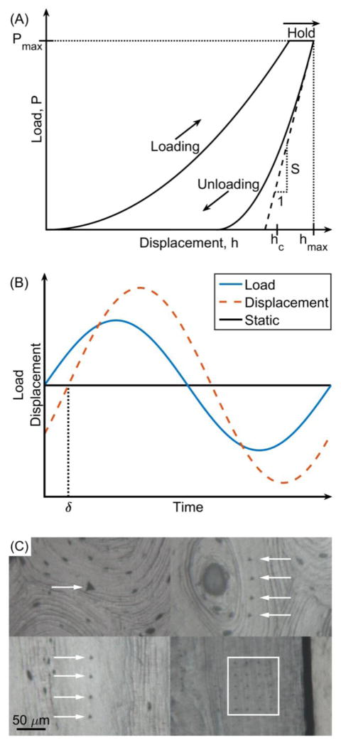
Tissue-level mechanical properties characterize mechanical behavior independently of microscopic porosity. Specifically, quasi-static nanoindentation provides measurements of modulus (stiffness) and hardness (resistance to yielding) of tissue at the length scale of the lamella, while dynamic nanoindentation assesses time-dependent behavior in the form of storage modulus (stiffness), loss modulus (dampening), and loss factor (ratio of the two). While these properties are useful in establishing how a gene, signaling pathway, or disease of interest affects bone tissue, they generally do not vary with aging after skeletal maturation or with osteoporosis. Heterogeneity in tissue-level mechanical properties or in compositional properties may contribute to fracture risk, but a consensus on whether the contribution is negative or positive has not emerged. In vivo indentation of bone tissue is now possible, and the mechanical resistance to microindentation has the potential for improving fracture risk assessment, though determinants are currently unknown.
Keywords: Bone quality; Bound water; Hardness; Nanoindentation; Spectroscopy; Viscoelasticity.
Figures
Similar articles
-
Modifications to nano- and microstructural quality and the effects on mechanical integrity in Paget's disease of bone.J Bone Miner Res. 2015 Feb;30(2):264-73. doi: 10.1002/jbmr.2340. J Bone Miner Res. 2015. PMID: 25112610
-
Increased tissue-level storage modulus and hardness with age in male cortical bone and its association with decreased fracture toughness.Bone. 2021 Jul;148:115949. doi: 10.1016/j.bone.2021.115949. Epub 2021 Apr 14. Bone. 2021. PMID: 33862261 Free PMC article.
-
Mechanical properties of cortical bone and their relationships with age, gender, composition and microindentation properties in the elderly.Bone. 2016 Dec;93:196-211. doi: 10.1016/j.bone.2015.11.018. Epub 2015 Dec 4. Bone. 2016. PMID: 26656135
-
On the Relation of Bone Mineral Density and the Elastic Modulus in Healthy and Pathologic Bone.Curr Osteoporos Rep. 2018 Aug;16(4):404-410. doi: 10.1007/s11914-018-0449-5. Curr Osteoporos Rep. 2018. PMID: 29869752 Review.
-
Bone strength in children: understanding basic bone biomechanics.Arch Dis Child Educ Pract Ed. 2016 Feb;101(1):2-7. doi: 10.1136/archdischild-2015-308597. Epub 2015 Aug 12. Arch Dis Child Educ Pract Ed. 2016. PMID: 26269494 Review.
Cited by
-
Effect of hormone replacement therapy on bone formation quality and mineralization regulation mechanisms in early postmenopausal women.Bone Rep. 2021 Mar 23;14:101055. doi: 10.1016/j.bonr.2021.101055. eCollection 2021 Jun. Bone Rep. 2021. PMID: 33850974 Free PMC article.
-
Non-Obese MKR Mouse Model of Type 2 Diabetes Reveals Skeletal Alterations in Mineralization and Material Properties.JBMR Plus. 2021 Dec 16;6(2):e10583. doi: 10.1002/jbm4.10583. eCollection 2022 Feb. JBMR Plus. 2021. PMID: 35229063 Free PMC article.
-
Bone intrinsic material and compositional properties in postmenopausal women diagnosed with long-term Type-1 diabetes.Bone. 2023 Sep;174:116832. doi: 10.1016/j.bone.2023.116832. Epub 2023 Jun 27. Bone. 2023. PMID: 37385427 Free PMC article.
-
Applying Full Spectrum Analysis to a Raman Spectroscopic Assessment of Fracture Toughness of Human Cortical Bone.Appl Spectrosc. 2017 Oct;71(10):2385-2394. doi: 10.1177/0003702817718149. Epub 2017 Jul 14. Appl Spectrosc. 2017. PMID: 28708001 Free PMC article.
-
Clinical measurements of bone tissue mechanical behavior using reference point indentation.Clin Rev Bone Miner Metab. 2018 Sep;16(3):87-94. doi: 10.1007/s12018-018-9249-9. Epub 2018 Sep 12. Clin Rev Bone Miner Metab. 2018. PMID: 30983912 Free PMC article.
References
-
- Lewis G, Nyman JS. The use of nanoindentation for characterizing the properties of mineralized hard tissues: state-of-the art review. Journal of biomedical materials research Part B, Applied biomaterials. 2008;87(1):286–301. - PubMed
-
- Thurner PJ. Atomic force microscopy and indentation force measurement of bone. Wiley Interdiscip Rev Nanomed Nanobiotechnol. 2009;1(6):624–49. - PubMed
-
- Liu XS, Stein EM, Zhou B, Zhang CA, Nickolas TL, Cohen A, et al. Individual trabecula segmentation (ITS)-based morphological analyses and microfinite element analysis of HR-pQCT images discriminate postmenopausal fragility fractures independent of DXA measurements. J Bone Miner Res. 2012;27(2):263–72. - PMC - PubMed
-
- Nishiyama KK, Macdonald HM, Hanley DA, Boyd SK. Women with previous fragility fractures can be classified based on bone microarchitecture and finite element analysis measured with HR-pQCT. Osteoporos Int. 2013;24(5):1733–40. - PubMed
Publication types
MeSH terms
Grants and funding
LinkOut - more resources
Full Text Sources
Other Literature Sources
Medical
