

Anatomical Connections of the Functionally Defined "Face Patches" in the Macaque Monkey
- PMID: 27263973
- PMCID: PMC5573145
- DOI: 10.1016/j.neuron.2016.05.009
Anatomical Connections of the Functionally Defined "Face Patches" in the Macaque Monkey
Abstract
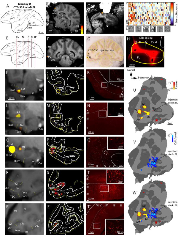
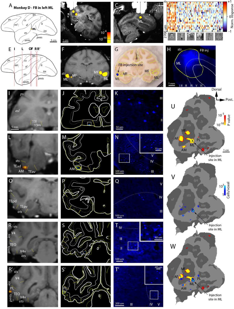
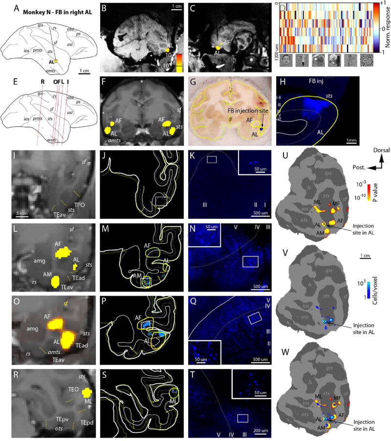
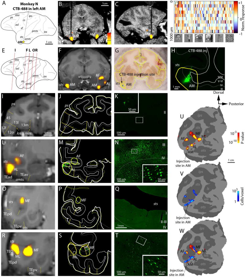
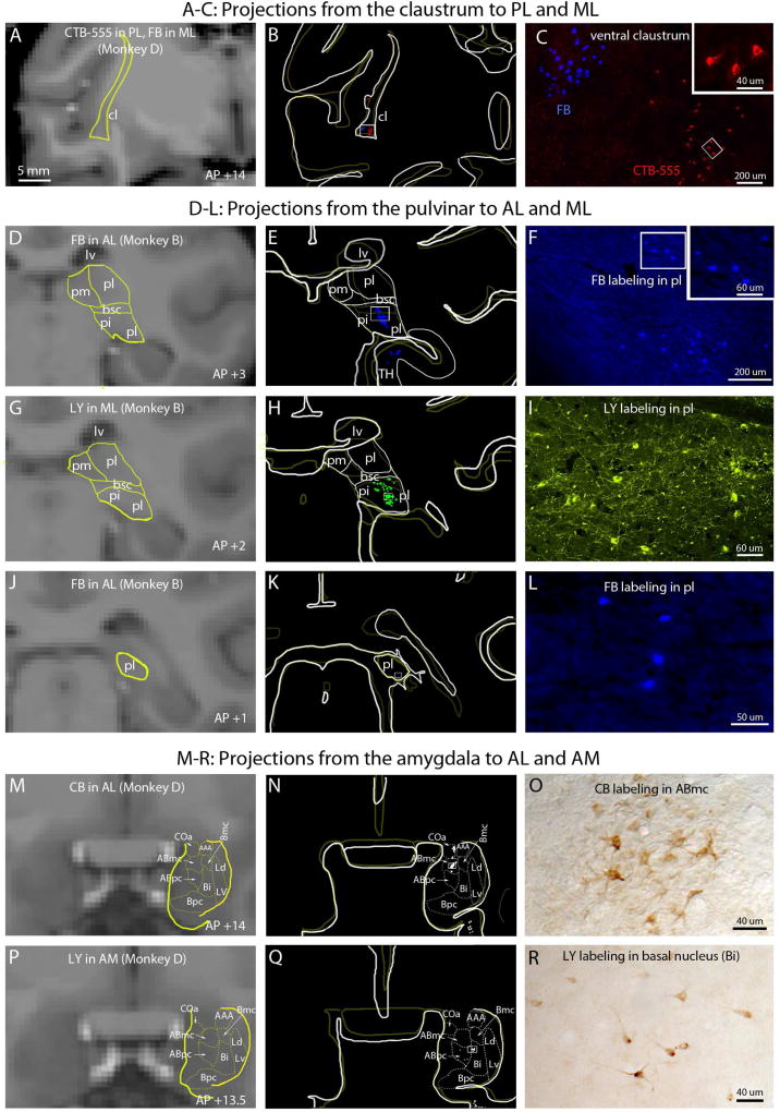
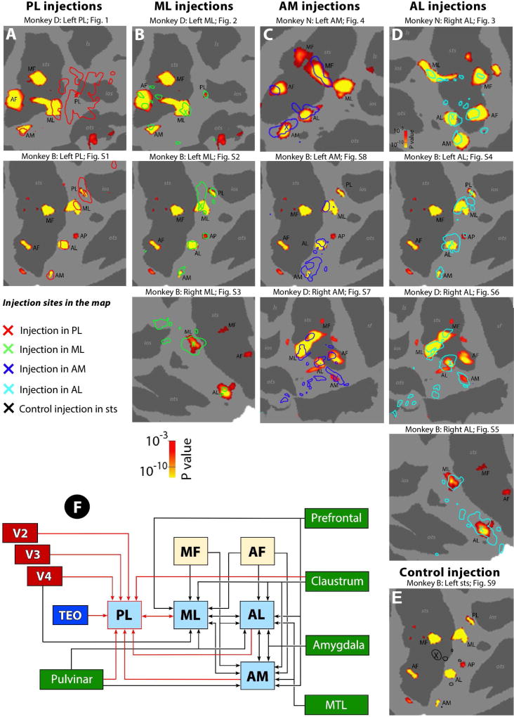
The neural circuits underlying face recognition provide a model for understanding visual object representation, social cognition, and hierarchical information processing. A fundamental piece of information lacking to date is the detailed anatomical connections of the face patches. Here, we injected retrograde tracers into four different face patches (PL, ML, AL, AM) to characterize their anatomical connectivity. We found that the patches are strongly and specifically connected to each other, and individual patches receive inputs from extrastriate cortex, the medial temporal lobe, and three subcortical structures (the pulvinar, claustrum, and amygdala). Inputs from prefrontal cortex were surprisingly weak. Patches were densely interconnected to one another in both feedforward and feedback directions, inconsistent with a serial hierarchy. These results provide the first direct anatomical evidence that the face patches constitute a highly specialized system and suggest that subcortical regions may play a vital role in routing face-related information to subsequent processing stages.
Copyright © 2016 Elsevier Inc. All rights reserved.
Figures
Similar articles
-
Pulvinar and other subcortical connections of dorsolateral visual cortex in monkeys.J Comp Neurol. 2002 Aug 26;450(3):215-40. doi: 10.1002/cne.10298. J Comp Neurol. 2002. PMID: 12209852
-
Subcortical connections of area V4 in the macaque.J Comp Neurol. 2014 Jun 1;522(8):1941-65. doi: 10.1002/cne.23513. J Comp Neurol. 2014. PMID: 24288173 Free PMC article.
-
Connections between the pulvinar complex and cytochrome oxidase-defined compartments in visual area V2 of macaque monkey.Exp Brain Res. 1995;104(3):419-30. doi: 10.1007/BF00231977. Exp Brain Res. 1995. PMID: 7589294
-
Corticocortical and thalamocortical information flow in the primate visual system.Prog Brain Res. 2005;149:173-85. doi: 10.1016/S0079-6123(05)49013-5. Prog Brain Res. 2005. PMID: 16226584 Review.
-
The Second Visual System of The Tree Shrew.J Comp Neurol. 2019 Feb 15;527(3):679-693. doi: 10.1002/cne.24413. Epub 2018 Mar 9. J Comp Neurol. 2019. PMID: 29446088 Free PMC article. Review.
Cited by
-
Advances in nonhuman primate models of autism: Integrating neuroscience and behavior.Exp Neurol. 2018 Jan;299(Pt A):252-265. doi: 10.1016/j.expneurol.2017.07.021. Epub 2017 Aug 1. Exp Neurol. 2018. PMID: 28774750 Free PMC article. Review.
-
A new no-report paradigm reveals that face cells encode both consciously perceived and suppressed stimuli.Elife. 2020 Nov 11;9:e58360. doi: 10.7554/eLife.58360. Elife. 2020. PMID: 33174836 Free PMC article.
-
Cortex Is Cortex: Ubiquitous Principles Drive Face-Domain Development.Trends Cogn Sci. 2019 Jan;23(1):3-4. doi: 10.1016/j.tics.2018.10.009. Epub 2018 Nov 24. Trends Cogn Sci. 2019. PMID: 30482446 Free PMC article. No abstract available.
-
On the relationship between maps and domains in inferotemporal cortex.Nat Rev Neurosci. 2021 Sep;22(9):573-583. doi: 10.1038/s41583-021-00490-4. Epub 2021 Aug 3. Nat Rev Neurosci. 2021. PMID: 34345018 Free PMC article. Review.
-
Audiovisual integration in macaque face patch neurons.Curr Biol. 2021 May 10;31(9):1826-1835.e3. doi: 10.1016/j.cub.2021.01.102. Epub 2021 Feb 25. Curr Biol. 2021. PMID: 33636119 Free PMC article.
References
-
- Baldauf D, Desimone R. Neural mechanisms of object-based attention. Science. 2014;344:424–427. - PubMed
-
- Cheng X, Crapse T, Tsao DY. Features that drive face cells: A comparison across face patches Paper presented at: Society for Neuroscience Conference; San Diego, CA. 2013.
MeSH terms
Grants and funding
LinkOut - more resources
Full Text Sources
Other Literature Sources
