Synchrotron X-ray microtransections: a non invasive approach for epileptic seizures arising from eloquent cortical areas
- PMID: 27264273
- PMCID: PMC4893707
- DOI: 10.1038/srep27250
Synchrotron X-ray microtransections: a non invasive approach for epileptic seizures arising from eloquent cortical areas
Abstract
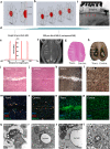
Synchrotron-generated X-ray (SRX) microbeams deposit high radiation doses to submillimetric targets whilst minimizing irradiation of neighboring healthy tissue. We developed a new radiosurgical method which demonstrably transects cortical brain tissue without affecting adjacent regions. We made such image-guided SRX microtransections in the left somatosensory cortex in a rat model of generalized epilepsy using high radiation doses (820 Gy) in thin (200 μm) parallel slices of tissue. This procedure, targeting the brain volume from which seizures arose, altered the abnormal neuronal activities for at least 9 weeks, as evidenced by a decrease of seizure power and coherence between tissue slices in comparison to the contralateral cortex. The brain tissue located between transections stayed histologically normal, while the irradiated micro-slices remained devoid of myelin and neurons two months after irradiation. This pre-clinical proof of concept highlights the translational potential of non-invasive SRX transections for treating epilepsies that are not eligible for resective surgery.
Figures


Similar articles
-
Synchrotron-generated microbeam sensorimotor cortex transections induce seizure control without disruption of neurological functions.PLoS One. 2013;8(1):e53549. doi: 10.1371/journal.pone.0053549. Epub 2013 Jan 14. PLoS One. 2013. PMID: 23341950 Free PMC article.
-
Synchrotron X-ray interlaced microbeams suppress paroxysmal oscillations in neuronal networks initiating generalized epilepsy.Neurobiol Dis. 2013 Mar;51:152-60. doi: 10.1016/j.nbd.2012.11.005. Epub 2012 Nov 16. Neurobiol Dis. 2013. PMID: 23159741
-
Microradiosurgical cortical transections generated by synchrotron radiation.Phys Med. 2015 Sep;31(6):642-6. doi: 10.1016/j.ejmp.2015.05.007. Epub 2015 May 29. Phys Med. 2015. PMID: 26032004
-
Synchrotron-generated microbeam radiosurgery: a novel experimental approach to modulate brain function.Neurol Res. 2011 Oct;33(8):825-31. doi: 10.1179/016164111X13123658647445. Neurol Res. 2011. PMID: 22004705 Review.
-
Microbeam radiosurgery using synchrotron-generated submillimetric beams: a new tool for the treatment of brain disorders.Neurosurg Rev. 2010 Apr;34(2):133-42. doi: 10.1007/s10143-010-0292-3. Epub 2010 Nov 19. Neurosurg Rev. 2010. PMID: 21088863 Review.
Cited by
-
Neuro-Oncologic Veterinary Trial for the Clinical Transfer of Microbeam Radiation Therapy: Acute to Subacute Radiotolerance after Brain Tumor Irradiation in Pet Dogs.Cancers (Basel). 2024 Jul 29;16(15):2701. doi: 10.3390/cancers16152701. Cancers (Basel). 2024. PMID: 39123429 Free PMC article.
-
Locomotion and eating behavior changes in Yucatan minipigs after unilateral radio-induced ablation of the caudate nucleus.Sci Rep. 2019 Nov 19;9(1):17082. doi: 10.1038/s41598-019-53518-2. Sci Rep. 2019. PMID: 31745153 Free PMC article.
-
In Vivo Femtosecond Laser Subsurface Cortical Microtransections Attenuate Acute Rat Focal Seizures.Cereb Cortex. 2019 Jul 22;29(8):3415-3426. doi: 10.1093/cercor/bhy210. Cereb Cortex. 2019. PMID: 30192931 Free PMC article.
-
Microbeam radiation therapy - grid therapy and beyond: a clinical perspective.Br J Radiol. 2017 Oct;90(1078):20170073. doi: 10.1259/bjr.20170073. Epub 2017 Jul 27. Br J Radiol. 2017. PMID: 28749174 Free PMC article. Review.
-
Synchrotron Radiation-Based Three-Dimensional Visualization of Angioarchitectural Remodeling in Hippocampus of Epileptic Rats.Neurosci Bull. 2020 Apr;36(4):333-345. doi: 10.1007/s12264-019-00450-0. Epub 2019 Dec 10. Neurosci Bull. 2020. PMID: 31823302 Free PMC article.
References
-
- Kahane P. & Bartolomei F. Temporal lobe epilepsy and hippocampal sclerosis: lessons from depth EEG recordings. Epilepsia 51 Suppl 1, 59–62 (2010). - PubMed
-
- Tellez-Zenteno J. F., Dhar R. & Wiebe S. Long-term seizure outcomes following epilepsy surgery: a systematic review and meta-analysis. Brain 128, 1188–1198 (2005). - PubMed
-
- Behdad A., Limbrick D. D., Bertrand M. E. & Smyth M. D. Epilepsy surgery in children with seizures arising from the rolandic cortex. Epilepsia 50, 1450–61 (2009). - PubMed
-
- Pondal-Sordo M., Diosy D., Téllez-Zenteno J. F., Girvin J. P. & Wiebe S. Epilepsy surgery involving the sensory-motor cortex. Brain 129, 3307–14 (2006). - PubMed
Publication types
MeSH terms
LinkOut - more resources
Full Text Sources
Other Literature Sources
Medical

