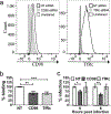
Select membrane proteins modulate MNV-1 infection of macrophages and dendritic cells in a cell type-specific manner
- PMID: 27264433
- PMCID: PMC6053272
- DOI: 10.1016/j.virusres.2016.06.001
Select membrane proteins modulate MNV-1 infection of macrophages and dendritic cells in a cell type-specific manner
Abstract
Noroviruses cause gastroenteritis in humans and other animals, are shed in the feces, and spread through the fecal-oral route. Host cellular expression of attachment and entry receptors for noroviruses is thought to be a key determinant of cell tropism and the strict species-specificity. However, to date, only carbohydrates have been identified as attachment receptors for noroviruses. Thus, we investigated whether host cellular proteins play a role during the early steps of norovirus infection. We used murine norovirus (MNV) as a representative norovirus, since MNV grows well in tissue culture and is a frequently used model to study basic aspects of norovirus biology. Virus overlay protein binding assay followed by tandem mass spectrometry analysis was performed in two permissive cell lines, RAW264.7 (murine macrophages) and SRDC (murine dendritic cells) to identify four cellular membrane proteins as candidates. Loss-of-function studies revealed that CD36 and CD44 promoted MNV-1 binding to primary dendritic cells, while CD98 heavy chain (CD98) and transferrin receptor 1 (TfRc) facilitated MNV-1 binding to RAW 264.7 cells. Furthermore, the VP1 protruding domain of MNV-1 interacted directly with the extracellular domains of recombinant murine CD36, CD98 and TfRc by ELISA. Additionally, MNV-1 infection of RAW 264.7 cells was enhanced by soluble rCD98 extracellular domain. These studies demonstrate that multiple membrane proteins can promote efficient MNV-1 infection in a cell type-specific manner. Future studies are needed to determine the molecular mechanisms by which each of these proteins affect the MNV-1 infectious cycle.
Keywords: CD36; CD44; CD98 heavy chain; Infection; Norovirus; Transferrin receptor 1.
Copyright © 2016 Elsevier B.V. All rights reserved.
Figures


Similar articles
-
Functional receptor molecules CD300lf and CD300ld within the CD300 family enable murine noroviruses to infect cells.Proc Natl Acad Sci U S A. 2016 Oct 11;113(41):E6248-E6255. doi: 10.1073/pnas.1605575113. Epub 2016 Sep 28. Proc Natl Acad Sci U S A. 2016. PMID: 27681626 Free PMC article.
-
Akt Plays Differential Roles during the Life Cycles of Acute and Persistent Murine Norovirus Strains in Macrophages.J Virol. 2022 Feb 9;96(3):e0192321. doi: 10.1128/JVI.01923-21. Epub 2021 Nov 17. J Virol. 2022. PMID: 34787460 Free PMC article.
-
Murine noroviruses bind glycolipid and glycoprotein attachment receptors in a strain-dependent manner.J Virol. 2012 May;86(10):5584-93. doi: 10.1128/JVI.06854-11. Epub 2012 Mar 21. J Virol. 2012. PMID: 22438544 Free PMC article.
-
Histologic Lesions Induced by Murine Norovirus Infection in Laboratory Mice.Vet Pathol. 2016 Jul;53(4):754-63. doi: 10.1177/0300985815618439. Epub 2016 Jan 20. Vet Pathol. 2016. PMID: 26792844 Free PMC article. Review.
-
Murine norovirus, a recently discovered and highly prevalent viral agent of mice.Lab Anim (NY). 2008 Jul;37(7):314-20. doi: 10.1038/laban0708-314. Lab Anim (NY). 2008. PMID: 18568010 Review.
Cited by
-
CD300LF Polymorphisms of Inbred Mouse Strains Confer Resistance to Murine Norovirus Infection in a Cell Type-Dependent Manner.J Virol. 2020 Aug 17;94(17):e00837-20. doi: 10.1128/JVI.00837-20. Print 2020 Aug 17. J Virol. 2020. PMID: 32581099 Free PMC article.
-
Functional receptor molecules CD300lf and CD300ld within the CD300 family enable murine noroviruses to infect cells.Proc Natl Acad Sci U S A. 2016 Oct 11;113(41):E6248-E6255. doi: 10.1073/pnas.1605575113. Epub 2016 Sep 28. Proc Natl Acad Sci U S A. 2016. PMID: 27681626 Free PMC article.
-
Bacterial extracellular vesicles control murine norovirus infection through modulation of antiviral immune responses.Front Immunol. 2022 Aug 4;13:909949. doi: 10.3389/fimmu.2022.909949. eCollection 2022. Front Immunol. 2022. PMID: 35990695 Free PMC article.
-
Current and Future Antiviral Strategies to Tackle Gastrointestinal Viral Infections.Microorganisms. 2021 Jul 27;9(8):1599. doi: 10.3390/microorganisms9081599. Microorganisms. 2021. PMID: 34442677 Free PMC article. Review.
-
Recent advances in understanding noroviruses.F1000Res. 2017 Jan 26;6:79. doi: 10.12688/f1000research.10081.1. eCollection 2017. F1000Res. 2017. PMID: 28163914 Free PMC article. Review.
References
-
- 2011Guide for the Care and Use of Laboratory Animals, 8th ed, Washington (DC).
-
- Aisen P, 2004. Transferrin receptor 1. The international journal of biochemistry & cell biology 36(11), 2137–2143. - PubMed
-
- Belliot G, Lopman BA, Ambert-Balay K, Pothier P, 2014. The burden of norovirus gastroenteritis: an important foodborne and healthcare-related infection. Clinical microbiology and infection : the official publication of the European Society of Clinical Microbiology and Infectious Diseases 20(8), 724–730. - PMC - PubMed
Publication types
MeSH terms
Substances
Grants and funding
LinkOut - more resources
Full Text Sources
Other Literature Sources
Medical
Miscellaneous

