. 2016 Jun 6:6:26960.
doi: 10.1038/srep26960.

Novel MRI Contrast Agent from Magnetotactic Bacteria Enables In Vivo Tracking of iPSC-derived Cardiomyocytes

Affiliations

Novel MRI Contrast Agent from Magnetotactic Bacteria Enables In Vivo Tracking of iPSC-derived Cardiomyocytes

Morteza Mahmoudi et al. Sci Rep. .

Abstract

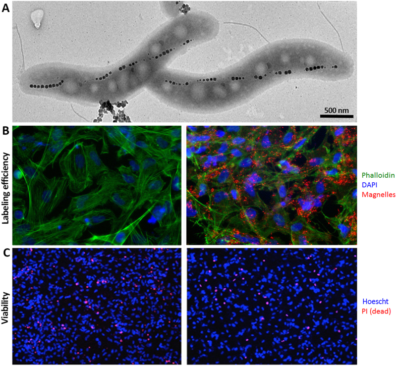
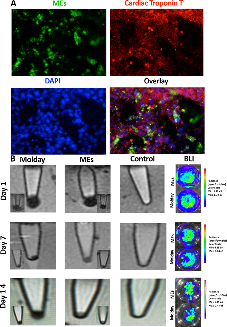
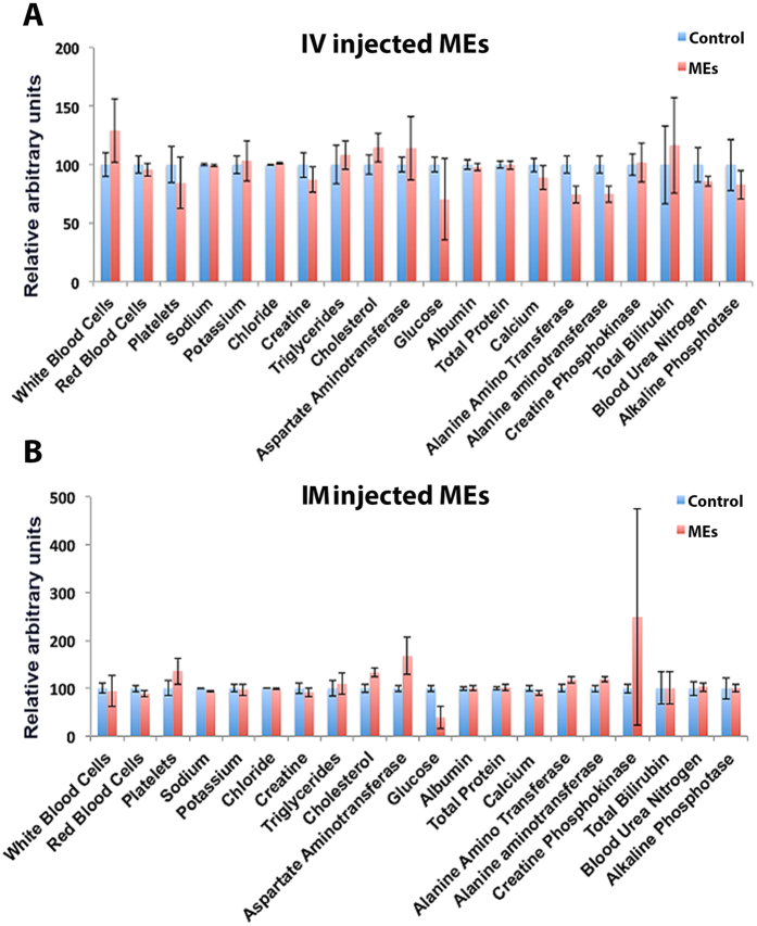
Therapeutic delivery of human induced pluripotent stem cell (iPSC)-derived cardiomyocytes (iCMs) represents a novel clinical approach to regenerate the injured myocardium. However, methods for robust and accurate in vivo monitoring of the iCMs are still lacking. Although superparamagnetic iron oxide nanoparticles (SPIOs) are recognized as a promising tool for in vivo tracking of stem cells using magnetic resonance imaging (MRI), their signal persists in the heart even weeks after the disappearance of the injected cells. This limitation highlights the inability of SPIOs to distinguish stem cell viability. In order to overcome this shortcoming, we demonstrate the use of a living contrast agent, magneto-endosymbionts (MEs) derived from magnetotactic bacteria for the labeling of iCMs. The ME-labeled iCMs were injected into the infarcted area of murine heart and probed by MRI and bioluminescence imaging (BLI). Our findings demonstrate that the MEs are robust and effective biological contrast agents to track iCMs in an in vivo murine model. We show that the MEs clear within one week of cell death whereas the SPIOs remain over 2 weeks after cell death. These findings will accelerate the clinical translation of in vivo MRI monitoring of transplanted stem cell at high spatial resolution and sensitivity.


Figures

Figure 1
Figure 1
(A) TEM image showing the magnetosome structure within MEs; (B) fluorescent images of the unlabeled (left panel) and ME labeled (right panel) iCMs stained with ME antibody in red, phalloidin in green and DAPI in blue, showing internalization of MEs in the iCMs; and (C) viability assessment of unlabeled (left panel) and ME labeled (right panel) iCMs using PI assay, dead cells are shown with a red signal while blue Hoescht staining corresponds to the total cell population.
Figure 2
Figure 2
(A) Fluorescent images of the ME-labeled iCMs used for the evaluation of potential ME effects on cardiac marker, ME staining in green, cardiac troponin T in red, and nuclei are counterstained with DAPI in blue and (B) MRI images (T2*) of the bottom of Eppendorf tubes of Molday ION labeled iCMs, ME-labeled iCMs, and PBS only control sample at different time points (days 1, 7, and 14 after labeling); the bottom panel shows the replicate MRI images for Molday ION and ME labeled cells. Right panel shows the corresponding BLI signal, indicating viability of the Molday ION and ME-labeled iCMs at days 1, 7, and 14 post labeling (the BLI experiments were performed in triplicate).
Figure 3
Figure 3
Relative concentrations liver and spleen enzymes and as well as key indices of infection and toxicity in blood did not changes in the highest doses of MEs (5.0E + 09) injected by both IV (A) and IM (B) routes.
Figure 4
Figure 4
Representative BLI images of the mice injected with ME and Molday ION labeled reporter gene-transduced iCMs or PBS control after (A) 7 days; (B) 14 days following cell injections (top) and their corresponding in vivo MRI images of the murine hearts (bottom). Red arrows show the signal from the injected labeled cells; (C) representative in vivo BLI images of the selected mice with dead cardiomyocytes at day 14 of ME labeled cells and the corresponding in vivo MRI images; (D) signal intensity (SI) for Molday ION (dark signal) and ME (with no signal). A significant absence of signal of ME* labeled dead cells compared to other samples with positive signal (p < 0.05).

References

    1. Urbanek K. et al.. Cardiac stem cells possess growth factor-receptor systems that after activation regenerate the infarcted myocardium, improving ventricular function and long-term survival. Circulation research 97, 663–673 (2005). - PubMed
    1. Urbanek K. et al.. Myocardial regeneration by activation of multipotent cardiac stem cells in ischemic heart failure. Proceedings of the National Academy of Sciences of the United States of America 102, 8692–8697 (2005). - PMC - PubMed
    1. van Berlo J. H. et al.. c-kit + cells minimally contribute cardiomyocytes to the heart. Nature 509, 337–341 (2014). - PMC - PubMed
    1. van Berlo J. H. & Molkentin J. D. An emerging consensus on cardiac regeneration. Nature medicine 20, 1386–1393 (2014). - PMC - PubMed
    1. Amir G. et al.. Dynamics of human myocardial progenitor cell populations in the neonatal period. The Annals of thoracic surgery 86, 1311–1319 (2008). - PubMed

LinkOut - more resources