. 2016 Jun 6:6:27459.
doi: 10.1038/srep27459.

IVA cloning: A single-tube universal cloning system exploiting bacterial In Vivo Assembly

Affiliations

IVA cloning: A single-tube universal cloning system exploiting bacterial In Vivo Assembly

Javier García-Nafría et al. Sci Rep. .

Abstract

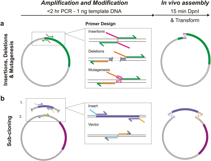
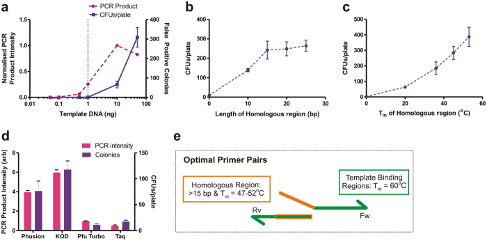
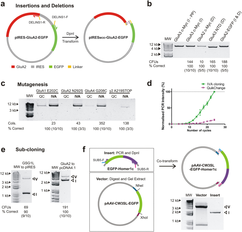
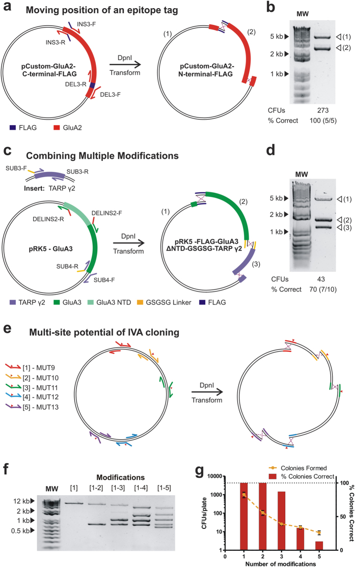
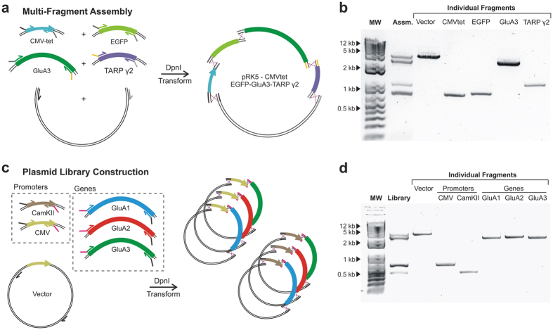
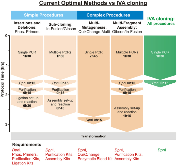
In vivo homologous recombination holds the potential for optimal molecular cloning, however, current strategies require specialised bacterial strains or laborious protocols. Here, we exploit a recA-independent recombination pathway, present in widespread laboratory E.coli strains, to develop IVA (In Vivo Assembly) cloning. This system eliminates the need for enzymatic assembly and reduces all molecular cloning procedures to a single-tube, single-step PCR, performed in <2 hours from setup to transformation. Unlike other methods, IVA is a complete system, and offers significant advantages over alternative methods for all cloning procedures (insertions, deletions, site-directed mutagenesis and sub-cloning). Significantly, IVA allows unprecedented simplification of complex cloning procedures: five simultaneous modifications of any kind, multi-fragment assembly and library construction are performed in approximately half the time of current protocols, still in a single-step fashion. This system is efficient, seamless and sequence-independent, and requires no special kits, enzymes or proprietary bacteria, which will allow its immediate adoption by the academic and industrial molecular biology community.


Figures

Figure 1
Figure 1. Method overview and primer design optimisation.
(a) Schematic of the universal IVA cloning protocol consisting of a single PCR reaction, producing homologous linear ends, followed by DpnI digestion and transformation, where amplified DNA is assembled in vivo by recombination. Primer design is shown for each type of basic modification: insertion, deletions, site-directed mutagenesis and sub-cloning. For insertions, the new sequence is best included in Fw and Rv primers, acting as the homologous region (magenta). For deletions, the overlap can be incorporated in any one primer, homologous to the other primer (orange) with primers straddling the undesired region (grey). Mutagenesis is similarly performed, inversely amplifying outside the undesired codon (ATG), with the replacement encoded in the forward primer (TGC). (b) Sub-cloning involves the amplification of both vector and insert in a single tube with homologous regions to directionally control assembly (blue and yellow).
Figure 2
Figure 2. Method and primer design optimisation.
(a) Performing PCR with no homologous regions highlights potential false positives arising from template DNA. Increasing template DNA increases the number of colonies produced on transformation (■ purple), independent of PCR amplification (● magenta). 1 ng regularly produces 0 colonies, yet gives substantial PCR amplification (dashed line). (b) Relationship between increasing length of homologous regions (constant Tm) and colony yield shows little length dependence of recombination above 15 bp. (c) Increasing the Tm of homologous regions increases the colony yield and hence recombination efficiency. (d) Bar chart indicates that IVA cloning colony yield (purple) is reliant on amount of PCR product (magenta) independent of the type of PCR polymerase. (e) Properties of optimum primer design to maximise recombination efficiency. Homologous regions are included in 5′-end of primers, homologous to a region (orange) of the partner primer. Template binding regions are shown in green.
Figure 3
Figure 3. Basic molecular cloning procedures using IVA cloning.
(a) Schematic depicting the simultaneous deletion of an IRES cassette (grey) and insertion of a linker sequence (yellow) in the GluA2-pIRES-EGFP vector. (b) Agarose gel showing the resulting amplification of insertions (I) and deletions (D). These include (Lane 2) insertion of a myc-tag at the N-terminus of GluA3 using phosphorylated primers (PP), and (Lane 3) IVA primers, (Lane 4) deletion of an N-terminal myc-tag in GluA2, (Lane 5) deletion of the N-terminal domain of GluA3 and (Lane 6) construction of a fusion GluA2-EGFP tandem construct by deleting the IRES cassette and introducing a linker. Number of colonies produced on transformation, and the percentage of colonies tested that contain the correct plasmid is shown below. MW  =  1 kb DNA ladder. (c) Agarose gel of PCR products providing a comparison between IVA and QuikChange TM mutagenesis primers. An enhancement of the intensity is seen for IVA primers in all cases. Number of colonies and percentage of correct clones for IVA cloning are shown below. (d) Cycle-by-cycle comparison of the PCR product formation between IVA (■ green) and QuikChangeTM (● magenta) for the GluA4 G208C mutation over 24 cycles of PCR (normalised to maximum value as 100%, n  =  3). The increased PCR yield of IVA is appreciable. (e) Agarose gel electrophoresis visualisation of PCR products for sub-cloning examples (GSG1L coding region into pIRES-mCherry and GluA2 coding region into pCDN4.1/TO) each showing two independent amplifications (Vector: V, Insert: I). Colony yields and percentage correct are shown below. (f) Alternative strategy for vectors not amenable to amplification, shown with the cloning of EGFP-Homer1c (Insert), subject to PCR, DpnI treatment and PCR purification, into the adeno-associated virus vector pAAV-CW3SL-EGFP (cut with NheI and XhoI, and gel purified. Agarose gel visualisation of vector post-digestion identifies gel purified fragment (V) alongside PCR amplified Insert (I).
Figure 4
Figure 4. One-tube multi-site modifications using IVA cloning.
(a) Schematic for multi-site modification whereby the position of a FLAG-tag (purple) is exchanged from the C- to the N-terminus of GluA2 (red) coding region in a CMV-based custom plasmid. The combination of deletion and insertion primers produces two amplification products after PCR. (b) The corresponding fragments (1 and 2) are visualised by agarose gel electrophoresis. (c) Schematic detailing multiple plasmid modification of GluA3-pRK5 vector in one tube. One set of primers a) deleted the N-terminal domain of GluA3 and b) inserted a FLAG-tag, while a second set of primers a) sub-cloned the TARP γ2 coding region (from a second vector) at the end of GluA3 and b) inserted a GSGSG linker to create a fusion construct. Together, these primers amplify three independent fragments, which are shown on an agarose gel (d). (d) Testing the number of multiple modifications that IVA cloning can perform simultaneously. Increasing number of XhoI restriction sites were created in the pRK5 plasmid using mutagenesis primers. Site of mutation is indicated by ▼. (f) PCR produced increasing numbers of bands corresponding to the number of modifications (1 to 1–5). (g) The number of colonies produced (yellow) and the percentage of correct clones (red) decreased with more modifications (n  =  3–5).
Figure 5
Figure 5. Single-tube multi-fragment assembly and library construction using IVA protocols.
(a) Schematic of a multi-fragment assembly where five independent fragments (CamKII, EGFP, GluA3 and TARP γ2 coding regions together with the pRK5 vector) were amplified in one PCR and assembled in vivo. (b) The amplification result is shown by agarose electrophoresis (Lane 2). Individual fragments were independently amplified to facilitate identification (Lanes 3–7). (c) Schematic of mammalian expression library construction. Two promotors (CamKII and CMV) and three genes (GluA1, GluA2 and GluA3 coding regions) where amplified in a single tube alongside the pRK5 vector. Assembly is guided by specific homologous regions that are shared within promotors and within genes. (d) Agarose electrophoresis resulting from the amplification in a single tube (Lane 2) with individual fragments shown (Lanes 3–8) to aid in the identification.
Figure 6
Figure 6. Comparing IVA with current optimal protocols for each cloning procedure.
Optimal methods for each type of cloning procedure have been selected (orange) for comparison with IVA (green). Labour time and requirements are shown for each example, with the universal IVA protocol significantly outperforming all methods, becoming the best option for all procedures. Of special importance are multisite applications (‘Complex Procedures’), where IVA halves the time required by the next best method and eliminates costs associated with enzymatic assembly and DNA. Furthermore, the IVA multi-site protocol surpasses optimal methods for performing single modifications (‘Simple Procedures’). All protocols require transformation into E. coli (grey). Contrasting with other methods, IVA only requires DpnI (‘Requirements’). (Phos.  =  phosphorylated).

References

    1. Green M. R. & Sambrook J. Molecular Cloning: A laboratory manual 4th edn, Vol. 1 Ch. 9, 157–260 (Cold Spring Harbour Laboratory Press, 2012).
    1. Kelwick R., MacDonald J. T., Webb A. J. & Freemont P. Developments in the Tools and Methodologies of Synthetic Biology. Front. Bioeng. Biotechnol. 2, 1–23 (2014). - PMC - PubMed
    1. Chen B.-Y. & Janes H. W. PCR Cloning Protocols in Methods in Molecular Biology 2nd edn, Vol. 192 (Humana Press, 2002).
    1. Mullis K. et al.. Specific enzymatic amplification of DNA in vitro: The polymerase chain reaction. Cold Spring Harb. Symp. Quant. Biol. 51, 263–273 (1986). - PubMed
    1. Bryksin A. & Matsamura I. Overlap Extension PCR Cloning. Methods Mol. Biol. 1073, 169–174 (2013). - PubMed

Publication types