. 2016 Jun 6:6:26979.
doi: 10.1038/srep26979.

The proteasome deubiquitinase inhibitor VLX1570 shows selectivity for ubiquitin-specific protease-14 and induces apoptosis of multiple myeloma cells

Affiliations

The proteasome deubiquitinase inhibitor VLX1570 shows selectivity for ubiquitin-specific protease-14 and induces apoptosis of multiple myeloma cells

Xin Wang et al. Sci Rep. .

Erratum in

Abstract

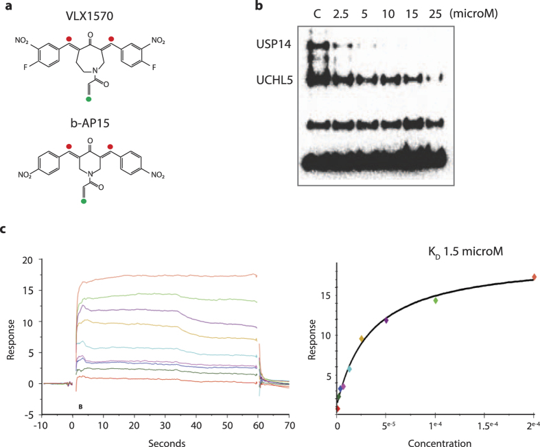
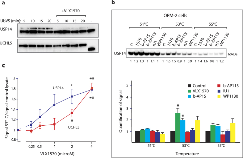
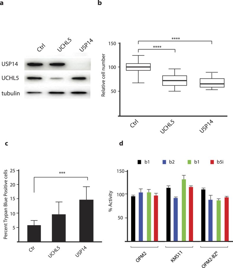
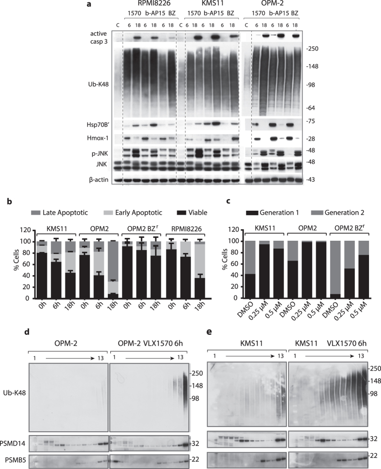
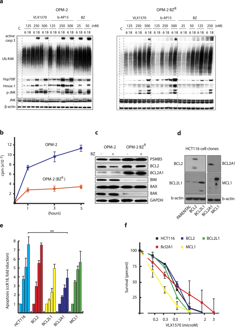
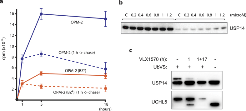
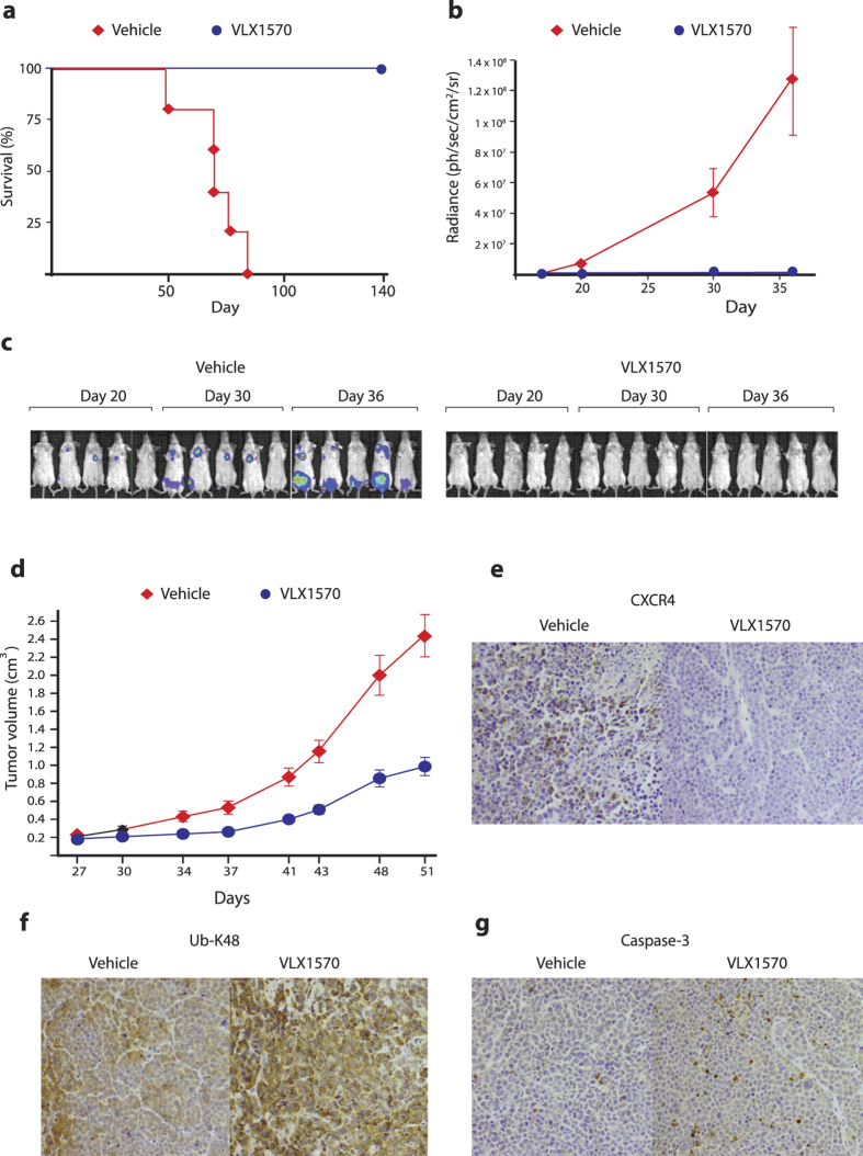
Inhibition of deubiquitinase (DUB) activity is a promising strategy for cancer therapy. VLX1570 is an inhibitor of proteasome DUB activity currently in clinical trials for relapsed multiple myeloma. Here we show that VLX1570 binds to and inhibits the activity of ubiquitin-specific protease-14 (USP14) in vitro, with comparatively weaker inhibitory activity towards UCHL5 (ubiquitin-C-terminal hydrolase-5). Exposure of multiple myeloma cells to VLX1570 resulted in thermostabilization of USP14 at therapeutically relevant concentrations. Transient knockdown of USP14 or UCHL5 expression by electroporation of siRNA reduced the viability of multiple myeloma cells. Treatment of multiple myeloma cells with VLX1570 induced the accumulation of proteasome-bound high molecular weight polyubiquitin conjugates and an apoptotic response. Sensitivity to VLX1570 was moderately affected by altered drug uptake, but was unaffected by overexpression of BCL2-family proteins or inhibitors of caspase activity. Finally, treatment with VLX1570 was found to lead to extended survival in xenograft models of multiple myeloma. Our findings demonstrate promising antiproliferative activity of VLX1570 in multiple myeloma, primarily associated with inhibition of USP14 activity.


Conflict of interest statement

J.G., P.D. and S.L. are shareholders in Vivolux AB.

Figures

Figure 1
Figure 1. VLX1570 inhibits and binds to proteasome DUBs in vitro.
(a) Structure of VLX1570 and b-AP15. The α,β-unsaturated carbonyls required for activity are marked with red filled circles and the Michael acceptor at the acrylamide is denoted with a green circle. (b) Inhibition of active-site-directed labeling of proteasomal deubiquitinases. Purified 19S proteasomes (5 nM) were pre-treated with DMSO or the indicated concentrations of VLX1570 (in DMSO) for 10 min at room temperature, followed by labeling with HA-Ub-VS and immunoblotting. (c) VLX1570 binding to USP14. Left: readings show binding signal in RU plotted against time in seconds. Higher concentrations clearly give rise to a higher binding signal. Right: points taken 10 seconds from the stop of injection in the reading to the left, plotted against the concentration on a linear scale. VLX1570 saturates the binding site in USP14 approaching a max signal and the fit of the data approaches the Rmax. The dissociation constant KD is read from the fit where the 1:1 binding isotherm has the value of 50% of the Rmax.
Figure 2
Figure 2. VLX1570 inhibits and binds to proteasome DUBs in exposed cells.
(a) Inhibition of active-site-directed labeling of proteasomal deubiquitinases by VLX1570 in MM cells. OPM-2 MM cells were exposed to 0.5 μM VLX1570 for 3 hours and 25 μg whole cell lysates were subsequently labeled with Ub-VS (1 μM), followed by SDS gel electrophoresis and immunoblotting with USP14 or UCHL5 antibodies. The upper bands represent active USP14 or UCHL5 enzymes. (b) Thermo-stabilization of USP14 in exposed cells. The same number of OPM-2 MM cells were exposed to DMSO, 1 μM VLX1570, 1 μM b-AP15, 10 μM b-AP113, 20 μM IU1 or 5 μM WP1130 for 1 hour and analysed for thermostability of UPS14 by CETSA (cellular thermal shift assay). The lower panel shows means ± S.E.M. for 3 different experiments (*p < 0.05 by t-test; calculated relative to the inactive control b-AP113). (c) Dose-response of thermal stabilization of USP14. OPM-2 myeloma cells were exposed to different concentrations of VLX1570 for 1 hour and collected for CETSA analysis. Shown are means ± S.E.M. for 3 different experiments (*p < 0.05; **p < 0.01 by t-test).
Figure 3
Figure 3. Knockdown of USP14 or UCHL5 results in loss of myeloma cell viability.
OPM-2 cells were transfected with siRNAs to USP14, UCHL5 or scrambled siRNAs. (a) Protein expression in transfected cells was determined using Western blotting after 72 hours; (b) the number of cells in different transfected cultures were determined. Shown are box-plots (median, quartiles and 10th and 90th percentiles). Differences were significant at the level of p < 0.0001 (Wilcoxon). (c) the number of dead cells was determined in transfected cultures 72 hours after transfection using Trypan Blue staining (p < 0.0005; Student’s t-test); (d) VLX1570 does not inhibit proteasome or immunoproteasome activity. OPM-2 cells were exposed to 0.5 μM VLX1570 for 3 hours and extracts were assayed using different proteasome substrates.
Figure 4
Figure 4. VLX1570 induces proteasome-associated polyubiquitin accumulation and apoptosis of multiple myeloma cells.
(a) MM cells were exposed to 0.5 μM VLX1570, 0.5 μM b-AP15 or 50 nM bortezomib (BZ) for 6 or 18 h, followed by immunoblotting for active caspase-3, Ub-K48, HSP70B  ´(HSPA6), Hmox-1, phospho-JNK, JNK and β-actin. RPMI8826 and KMS11 data are from the same filter, OPM-2 from a separate (dashed lines were introduced for clarity). (b) Determination of cell death induction after exposure to 0.5 μM VLX1570 or vehicle for 0, 6 or 18 h. Cells were stained with FITC-conjugated annexin V and propidium iodide and processed by flow cytometry. The percentage of viable, early apoptotic and late apoptotic were quantified. Results shown are mean values of triplicate measurements. (c) Estimation of cell division based on dilution of carboxyfluorescein succinimidyl ester (CFSE) membrane staining during cell division. Cells were exposed to 0.25 μM or 0.5 μM VLX1570 or DMSO as a control for 72 h. The fraction of cells in different generations was determined by flow cytometry analysis. (d,e) Analysis of the effects of VLX1570 on the sedimentation profiles of polyubiquitinated proteins. Cell lysates from control or VLX1570-exposed OPM-2 (c) or KMS-11 cells (d) were subjected to glycerol gradient centrifugation. Gradient fractions were collected (1 = top; 13 = bottom) and subjected to immunoblotting using antibodies to Ub-K48, PSMD14 (19S subunit), or PSMB5 (20S subunit). Note the accumulation of polyubiquitin on 26S proteasomes after 6 hours of drug exposure.
Figure 5
Figure 5. Examination of mechanisms affecting cellular sensitivity to VLX1570.
(a) Characterization of the bortezomib-resistant myeloma cell line OPM-BZR. Cells were exposed to VLX1570, b-AP15 or bortezomib (BZ) at the indicated concentrations for 6 or 18 h, followed by immunoblotting with Ub-K48, Hmox-1, HSP70B′, active-caspase-3, phospho-JNK, JNK, or β-actin antibodies. OPM-2 and OPM-2BZR samples are from separate filters, for comparision on the same filters see Supplementary Fig. 5d; (b) Uptake of 3H-VLX1570 by OPM-2 and OPM-2-BZR cells. Cells were incubated with labeled drug in growth medium at 37 °C, washed and processed for liquid scintillation counting. (c) Expression of apoptotic regulators and PSMB5 in OPM-2 and OPM-2 BZR cells. Note that long-term selection in the presence of bortezomib resulted in altered expression of BCL2, BCL2A1, BIM and BAK but that the expression of these proteins is not affected by acute drug exposure. PSMB5 is a 20S proteasome subunit, the expression of which is known to be associated with bortezomib resistance. (d) Expression of BCL2-family proteins in HCT116 cell clones (full images are shown in Supplementary Fig. 8); (e) Apoptosis induction of HCT116 cells infected with lentiviruses expressing different BCL2 family members following exposure to 1 μM VLX1570 for 18 hours. Accumulation of caspase-cleaved K18 fragments in cells and culture media was measured by ELISA. (f) Survival of HCT116 cells infected with lentiviruses expressing different BCL22 family members and exposed to different concentrations μM VLX1570. Survival was measured at 48 hours.
Figure 6
Figure 6. Retention of VLX1570 and target engagement after drug wash-out.
(a) OPM-2 and OPM-2-BZR cells were exposed to VLX1570 for 1 hour and the uptake of 3H-VLX1570 was determined immediately or after an additional 17 hours incubation in drug-free medium; (b) Thermostabilization of USP14 was determined 17 hours after wash-out of VLX1570 by CETSA. Equal number of cells were used at each time point (loading controls are not relevant in this type of analysis23), (c) DUB inhibition was determined by Ub-VS labeling of cell extracts after 1 h exposure to VLX1570 and after 1 h exposure followed by 17 hours wash-out.
Figure 7
Figure 7. In vivo activity of VLX1570 in multiple myeloma xenografts.
(a) KMS-11-LUC2 cells (5 × 106) were injected intravenously into female SCID mice. After 7 days, mice were randomized into control and treatment groups (5 mice per group). Mice were treated with vehicle or VLX1570 (3 mg/kg) for 10 consecutive days. The drug was dissolved in PEG/Chremophore/Tween (50/10/40) and diluted 1:10 with saline prior to intravenous injection. Shown is survival over a 142 day period. (b) Quantification of bioluminescence measured at 18, 20, and 30 and 36 days of tumor cell injection. (c) Bioluminescence recorded in individual animals. (d) Growth of subcutaneous RPMI8226 tumors following treatment with VLX1570. Mice were exposed to VLX1570 dissolved in PEG/Chremophore/Tween (50/10/40). Mice were treated with 3 mg/kg VLX1570 for 10 consecutive days (5 mice per group). (e) Immunohistochemical staining showing decreased levels of CXCR4 in RPMI8226 tumors after exposure to 3 mg/kg VLX1570. (f) Increased immunohistochemical staining of K48-linked ubiquitin chains in RPMI8226 tumors after exposure to 3 mg/kg VLX1570. (g) Increased caspase-3 activity after exposure of RPMI8226 tumors to 3 mg/kg VLX1570.

Similar articles

Cited by

References

    1. Hershko A. & Ciechanover A. The ubiquitin system. Ann Rev Biochem 67, 425–479 (1998). - PubMed
    1. Groll M. et al. Structure of 20S proteasome from yeast at 2.4 A resolution. Nature 386, 463–471 (1997). - PubMed
    1. Goldberg A. L. Functions of the proteasome: from protein degradation and immune surveillance to cancer therapy. Biochem Soc Trans 35, 12–17 (2007). - PubMed
    1. Groll M. et al. The catalytic sites of 20S proteasomes and their role in subunit maturation: a mutational and crystallographic study. Proc Natl Acad Sci USA 96, 10976–10983 (1999). - PMC - PubMed
    1. Lee M. J., Lee B. H., Hanna J., King R. W. & Finley D. Trimming of ubiquitin chains by proteasome-associated deubiquitinating enzymes. Mol Cell Proteomics 10, R110 003871 (2011). - PMC - PubMed

Publication types

MeSH terms