Review
. 2016 Oct;196(1):3-14.
doi: 10.1016/j.jsb.2016.05.010. Epub 2016 Jun 2.

Membrane remodelling in bacteria

Affiliations
Review

Membrane remodelling in bacteria

Olga Bohuszewicz et al. J Struct Biol. 2016 Oct.

Abstract

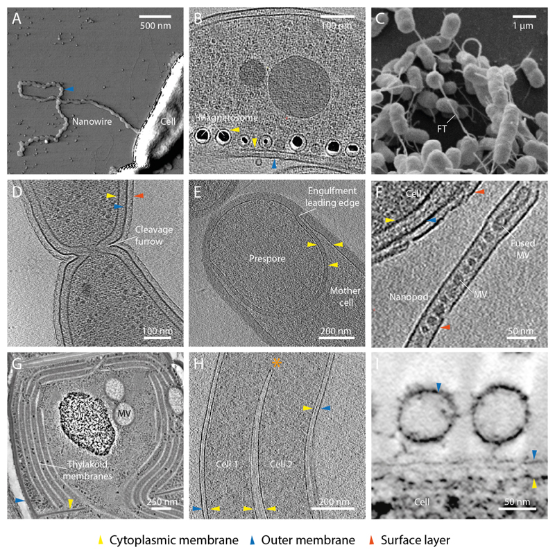
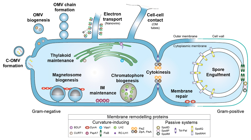
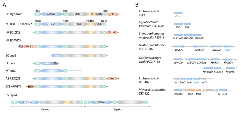
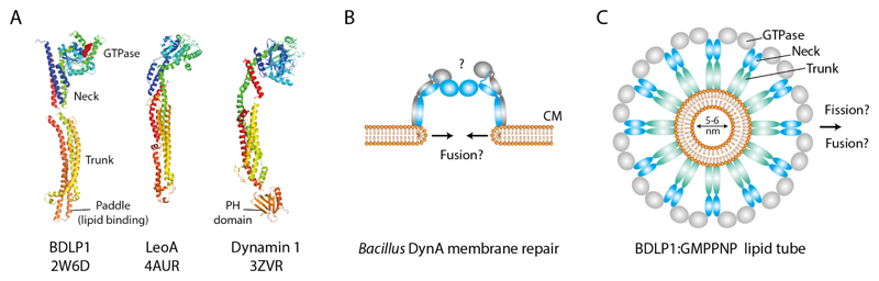
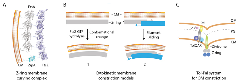
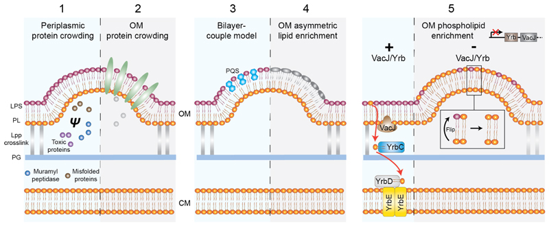
In bacteria the ability to remodel membrane underpins basic cell processes such as growth, and more sophisticated adaptations like inter-cell crosstalk, organelle specialisation, and pathogenesis. Here, selected examples of membrane remodelling in bacteria are presented and the diverse mechanisms for inducing membrane fission, fusion, and curvature discussed. Compared to eukaryotes, relatively few curvature-inducing proteins have been characterised so far. Whilst it is likely that many such proteins remain to be discovered, it also reflects the importance of alternative membrane remodelling strategies in bacteria where passive mechanisms for generating curvature are utilised.

Keywords: Bacteria; Curvature-inducing protein; Membrane; Remodelling; Vesicle.


Figures

Fig. 1
Fig. 1
Selected examples of high membrane curvature and remodelling in bacteria. (A) Atomic force microscopy image of a Shewanella oniedensis MR-1 cell with an OM extension that forms a nanowire (Pirbadian et al., 2014). Image courtesy of Sahand Pirbadian and Mohamed El-Naggar. (B) Electron cryo tomogram (ECT) of Magnetospirillum magneticum AMB-1 shows that magnetosomes are invaginations of the CM (Komeili et al., 2006). Image courtesy of Arash Komeili and Grant Jensen. (C) Scanning electron micrograph showing cross-feeding between Escherichia coli and Acinetobacter baylyi connected by nanotubular membrane structures (Pande et al., 2015). FT- feeding tube. Image courtesy of Christian Kost. (D) ECT showing a late stage dividing Caulobacter crescentus cell with invaginated cytokinetic cleavage furrow. Image courtesy of Tanmay Bharat and Jan Löwe. (E) ECT of sporulating Bacillus subtilis. The prespore is in late stage engulfment by the mother cell (Tocheva et al., 2013). Image courtesy of Elitza Tocheva and Grant Jensen. (F) ECT of a Delftia sp. Cs1-4 nanopod (Shetty et al., 2011). MV- membrane vesicle. Image courtesy of Elitza Tocheva, Grant Jensen and William Hickey. (G) Thin-section micrograph of a high-pressure frozen, freeze-substituted Microcoleus sp. cell showing CM vesicles (MV) and extensive thylakoid membrane network (Scheuring et al., 2014). Image courtesy of Dana Charuvi, Reinat Nevo and Ziv Reich. (H) ECT of two Borrelia garinii cells with fused cell envelopes (Kudryashev et al., 2011). Asterisk shows merging of cytoplasmic cylinders and a region of high IM curvature. Image courtesy of Misha Kudryashev and Friedrich Frischknecht. (I) Micrograph of high pressure frozen, freeze substituted Myxococcus xanthus biofilms depicting OM vesicles tethered to cell surface (Palsdottir et al., 2009; Remis et al., 2014). Image courtesy of Manfred Auer.
Fig. 2
Fig. 2
Schematic showing key membrane remodelling events in bacteria. Proteins involved in membrane remodelling are divided into two categories depending on their ability to directly bind membrane and induce its curvature. Note that whilst BDLP1 is known to associate with the cell envelope, its role in thylakoid maintenance is currently only speculative. PspA is included as it senses and binds stored curvature elastic stress in the membrane (McDonald et al., 2015) but direct evidence that it is able to remodel membrane like its homologue Vipp1 is still lacking.
Fig. 3
Fig. 3
DFM gene structure and gene copy number is variable in bacteria. (A) Schematic showing the conserved modular structure of human Dynamin 1 (Faelber et al., 2011; Ford et al., 2011) and selected bacterial DFMs. Many bacterial DFMs have additional non-canonical domains that likely mediate protein-protein interactions. PRD- proline-rich domain; DnaJ- chaperone protein domain, PF00226; TM- predicted trans-membrane; TerB- tellurite resistance protein domain, PF05099; Ank- ankyrin repeat, PF12796; HTH- helix-turn-helix, PF01381. HS- Homo sapiens; NP- Nostoc punctiforme PCC73102; EC- Escherichia coli H10407; MT- Mycobacterium tuberculosis H37Rv; AS- Alistipes sp. CAG:435; ON- Oscillatoria nigro-viridis PCC 7112. (B) Schematic showing the variable number and genetic arrangement of bacterial DFMs between selected species (blue arrows). Bacterial DFMs usually exist as at least a pair in an operon (Bürmann et al., 2011; Michie et al., 2014). E. coli is an exception with the single DFM crfC (Ozaki et al., 2013). Cyanobacteria such as Nostoc punctiforme and Oscillatoria nigro-viridis usually have between 4-8 DFMs. Crystal structures for BDLP1 (Low and Löwe, 2006) and LeoA (Michie et al., 2014) confirm that their respective genes code for DFMs, whilst all others are assigned by sequence homology. In Myxococcus xanthus, two predicted bacterial DFMs flank the genes traA and traB, which are essential for OM exchange (Ducret et al., 2013; Pathak et al., 2012).
Fig. 4
Fig. 4
DFM-mediated membrane remodeling mechanisms in bacteria. (A) Bacterial and human DFMs share a conserved structural fold. (B) Bacillus subtilis DynA is implicated in membrane repair through a poorly understood fusion mechanism that appears not to rely on helical self-assembly as for BDLP1. (C) End-on view schematic showing a molecular model of a BDLP1-lipid tube (Low et al., 2009). BDLP1-GMPPNP forms a helical scaffold that forces the bound membrane into a highly curved tube. Through protein crowding and wedging, the paddle at the trunk tip likely displaces much of the outer leaflet.
Fig. 5
Fig. 5
Overview of core cytokinetic CM and OM remodeling machinery. (A) The proteins FtsA, ZipA and FtsZ form the Z-ring, a helical membrane-associated contractile scaffold that localises at the mid-cell. Minimally, FtsA and ZipA tether FtsZ to the membrane. FtsZ is the motor for generating membrane contractile force and curvature. (B) Two prevailing mechanisms for how FtsZ generates membrane curvature. Left, GTP hydrolysis drives FtsZ filament curving which is coupled to membrane bending. Right, Helical FtsZ filaments maximize lateral interactions between neighbouring rungs through filament sliding. This leads to constriction of the helix and the associated membrane. Both mechanisms require iterative cycles of FtsZ assembly and disassembly. (C) Schematic showing the core components of the Tol-Pal complex, which couples the CM to the OM, and is essential for OM constriction. A proton motive force is somehow required for energisation and function of the complex.
Fig. 6
Fig. 6
Mechanisms of OMV biogenesis. All models depend on an initial decoupling of the OM from the peptidoglycan layer by breakage of the Lpp crosslink. This decoupling alone is sufficient to induce membrane vesiculation and can be augmented by the following 5 membrane curving mechanisms none of which involve CIPs (Schwechheimer and Kuehn, 2015). 1, Selective local protein crowding in the periplasm increases turgor pressure (ψ) and induces membrane bulging. 2, Selective crowding of cargo proteins such as virulence factors in the OM. 3, A generalised model for OMV formation where molecules such as Pseudomonas quinolone sequence (PQS) increase the surface area of the outer leaflet relative to the inner leaflet by a wedging effect (Schertzer and Whiteley, 2012). 4, Specific enrichment of different fatty acids and LPS subtypes that may be further modified by charge or polysaccharide incorporation. 5, A novel potentially general mechanism for OMV biogenesis based on phospholipid accumulation in the OM outer leaflet. In the absence of the VacJ/YrbABC transport system, phospholipids flip from the OM inner leaflet and are retained in the outer leaflet rather than being recovered to the CM (Roier et al., 2016).

References

    1. Allan EJ, Hoischen C, Gumpert J. Bacterial L-forms. Advances in applied microbiology. 2009;68:1–39. - PubMed
    1. Anderson DE, Gueiros-Filho FJ, Erickson HP. Assembly dynamics of FtsZ rings in Bacillus subtilis and Escherichia coli and effects of FtsZ-regulating proteins. J Bacteriol. 2004;186:5775–5781. - PMC - PubMed
    1. Armbruster U, Labs M, Pribil M, Viola S, Xu W, Scharfenberg M, Hertle AP, Rojahn U, Jensen PE, Rappaport F, Joliot P, et al. Arabidopsis CURVATURE THYLAKOID1 proteins modify thylakoid architecture by inducing membrane curvature. The Plant cell. 2013;25:2661–2678. - PMC - PubMed
    1. Berleman J, Auer M. The role of bacterial outer membrane vesicles for intra- and interspecies delivery. Environmental microbiology. 2013;15:347–354. - PubMed
    1. Bernhardt TG, de Boer PA. SlmA, a nucleoid-associated, FtsZ binding protein required for blocking septal ring assembly over chromosomes in E. coli. Molecular cell. 2005;18:555–564. - PMC - PubMed

Publication types

MeSH terms

Substances