. 2016 Aug 1;416(1):136-148.
doi: 10.1016/j.ydbio.2016.05.035. Epub 2016 Jun 2.

Pharyngeal morphogenesis requires fras1-itga8-dependent epithelial-mesenchymal interaction

Affiliations

Pharyngeal morphogenesis requires fras1-itga8-dependent epithelial-mesenchymal interaction

Jared Coffin Talbot et al. Dev Biol. .

Abstract

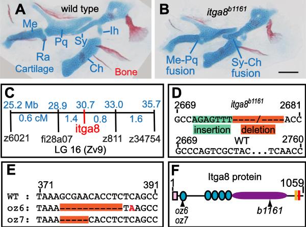
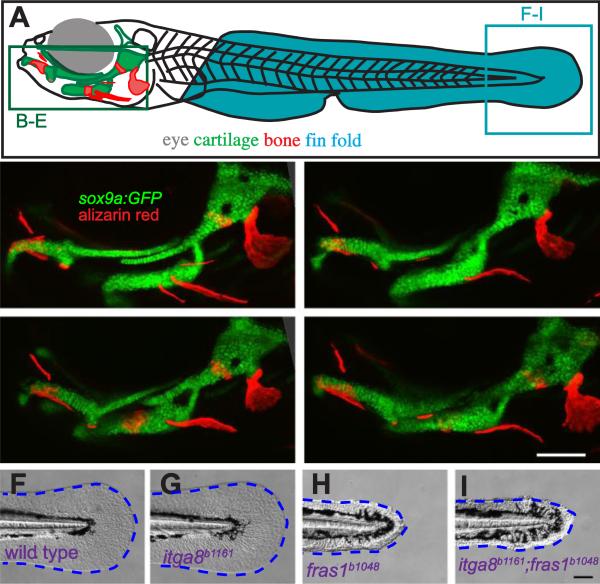
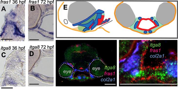
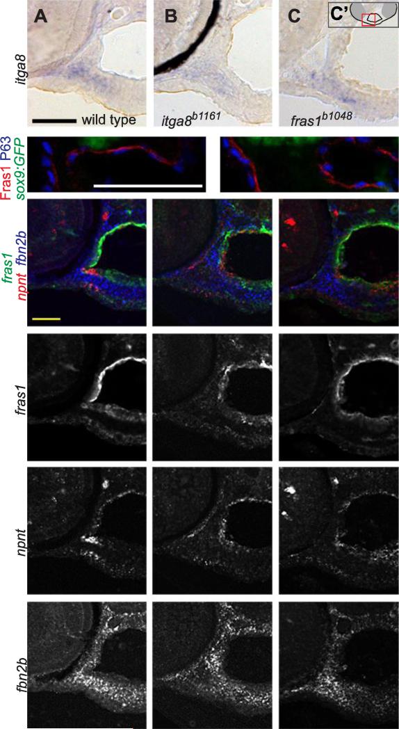
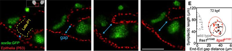
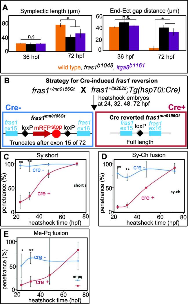
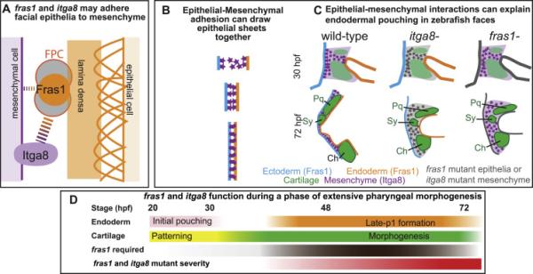
Both Fras1 and Itga8 connect mesenchymal cells to epithelia by way of an extracellular 'Fraser protein complex' that functions in signaling and adhesion; these proteins are vital to the development of several vertebrate organs. We previously found that zebrafish fras1 mutants have craniofacial defects, specifically, shortened symplectic cartilages and cartilage fusions that spare joint elements. During a forward mutagenesis screen, we identified a new zebrafish mutation, b1161, that we show here disrupts itga8, as confirmed using CRISPR-generated itga8 alleles. fras1 and itga8 single mutants and double mutants have similar craniofacial phenotypes, a result expected if loss of either gene disrupts function of the Fraser protein complex. Unlike fras1 mutants or other Fraser-related mutants, itga8 mutants do not show blistered tail fins. Thus, the function of the Fraser complex differs in the craniofacial skeleton and the tail fin. Focusing on the face, we find that itga8 mutants consistently show defective outpocketing of a late-forming portion of the first pharyngeal pouch, and variably express skeletal defects, matching previously characterized fras1 mutant phenotypes. In itga8 and fras1 mutants, skeletal severity varies markedly between sides, indicating that both mutants have increased developmental instability. Whereas fras1 is expressed in epithelia, we show that itga8 is expressed complementarily in facial mesenchyme. Paired with the observed phenotypic similarity, this expression indicates that the genes function in epithelial-mesenchymal interactions. Similar interactions between Fras1 and Itga8 have previously been found in mouse kidney, where these genes both regulate Nephronectin (Npnt) protein abundance. We find that zebrafish facial tissues express both npnt and the Fraser gene fibrillin2b (fbn2b), but their transcript levels do not depend on fras1 or itga8 function. Using a revertible fras1 allele, we find that the critical window for fras1 function in the craniofacial skeleton is between 1.5 and 3 days post fertilization, which coincides with the onset of fras1-dependent and itga8-dependent morphogenesis. We propose a model wherein Fras1 and Itga8 interact during late pharyngeal pouch morphogenesis to sculpt pharyngeal arches through epithelial-mesenchymal interactions, thereby stabilizing the developing craniofacial skeleton.


Figures

Figure 1
Figure 1
Skeletal defects in b1161 mutants are caused by lesions in itga8. (A, B) Alcian and alizarin staining of (A) wild type and (B) itga8b1161 homozygotes shows skeletal defects caused by itga8 mutations. Homozygous itga8 mutants often show cartilage fusions in the first two pharyngeal arches and defects in symplectic length. (C) Linkage analysis reveals no recombinants (0/696 individuals) between b1161 and the itga8b1161, placing them at the same map position. Map distances (blue) of additional markers from b1161 are shown above in Mb and below in cM. (D) Sequence of the itga8b1161 lesion reveals a 7 bp insertion in exon 25 (green), followed by a 79 bp deletion (orange) (wild type: GenBank JN399198, b1161: GenBank JN399198). This lesion results in protein truncation after amino acid 845 of 1059 with the predicted addition of 21 aberrant amino acids (EFTHWSWRPRLFRTLQSYWAS). (E) Sequence of two CRISPR-induced itga8 lesions, itga8oz6 (11 bp deletion) and itga8oz7 (5 bp deletion), reveal that both introduce frameshifts after amino acid 79 of 1059; itga8oz6 causes an immediate stop codon after the frameshift, while itga8oz7 introduces 30 aberrant amino acids (HLSAGDCGGRSGVLLPLAGIRPRLLPPDPL) before terminating. (F) Itga8 protein diagram with locations of oz6, oz7, and b1161 mutations along with predicted protein motifs. Protein motifs are designated as follows: signal sequence (pink box), integrin beta domains (teal circles); integrin alpha domain (purple oval), transmembrane domain (peach box), and an intracellular integrin domain (red box). Cartilage abbreviations: Meckel's (Me), Retroarticular process (Ra), Palatoquadrate (Pq), Symplectic (Sy), Ceratohyal (Ch), Interhyal (Ih). Scale bar (100 μm) in B also applies to A.
Figure 2
Figure 2
Comparison of skeletal, endodermal, and tail ectoderm phenotypes between wild type, itga8b1161 mutants, fras1b1048 mutants, and itga8b1161;fras1b1048 double mutants. (A) Illustration of a zebrafish larva, indicating regions shown in subsequent (B-I) panels. (B-E) Skeletal morphology is revealed using sox9a:EGFP expression (cartilage) and alizarin red staining (bone) at 7 dpf; itga8 and fras1 single and double mutants display similar cartilage defects, in particular, Meckel's-palatoquadrate joint fusion (arrowhead) and symplectic-ceratohyal fusions (asterisk). (F-I) Bright field images showing normal fin fold morphology (outlined blue) in (F) wild type and (G) itga8b1161 individuals versus the “blister phenotypes” in (H) fras1b1048 and (I) itga8b1161;fras1b1048 mutants. Scale bars (E, I) are 100 μm. Scale bar in E applies to B-E; Scale bar in I applies to F-I.
Figure 3
Figure 3
Phenotypic variation in fras1 and itga8 mutant phenotypes shows fluctuating asymmetry. (A-C’) Facial cartilage skeleton marked by sox9a:GFP expression at 7.5 dpf, region shown is boxed in red in A”. Compared to wild type (A), the itga8b1161 mutant skeleton is often asymmetric (B, C). For instance, the fish in (B) shows an extended symplectic cartilage fused to the ceratohyal cartilage on the right side and an unfused, severely shortened, symplectic phenotype on its left side (B’). In another example, both “Short Sy” and “Fused Sy-Ch” symplectic phenotypes (C) are found in a fish presenting only subtle defects on the opposite side (C’). (D, E) At 7.5 dpf, average symplectic length in itga8 mutants is shorter than wild type, but comparable to fras1 mutants. Symplectic cartilages in itga8 mutants show asymmetry similar to fras1 mutants, which is twice as high as wild-type asymmetry. (E) Plot of symplectic lengths measured on left and right sides, with grouped 95% density ellipses. Symplectic lengths for wild types were along the diagonal as expected for a high degree of left/right correlation, but for itga8b1161 and fras1b1048 mutants, symplectics were much shorter and do not correlate well between sides. Scale bar (A) is 100 μm, applicable to A-C’. Error bars (D) show 95% confidence intervals: 1.95 times standard error.
Figure 4
Figure 4
fras1 and itga8 are expressed in adjacent facial tissues. (A-D) Colorimetric in situ hybridization, developed using NBT/BCIP on wild-type sections, showing fras1 expression in pharyngeal and ectodermal epithelia (A, B) and itga8 expression in mesenchyme (C, D) at 36 hpf (A, C) and 72 hpf (B,D). (E) Color-coded diagram of lateral and transverse sections from a wild-type 60 hpf embryo, showing the first two pharyngeal arches (green), ectoderm (orange), cartilage (blue), and endoderm (red). (F, G) Fluorescent RNA in situ for fras1, itga8, and col2a1 expression in a 60 hpf transverse section of wild-type embryos at (F) low and (G) higher magnification. All scale bars: 50 μm.
Figure 5
Figure 5
fras1 and itga8 do not regulate one another, nor do they regulate candidate targets npnt or fbn2b. (A-C) RNA in situ hybridization for itga8 transcripts on transverse sections in 60 hpf wild-type and mutant embryos, developed colorimetrically, oriented as shown in C'. These transcripts appear similar in (A) wild type, (B) itga8 mutants, and (C) fras1 mutants. The black crescent in (B) is eye pigment. (D, E) 72 hpf tissue sections, labeled for Fras1 protein, epithelial nuclei (anti-P63) and cartilage (sox9a:GFP). Fras1 protein levels and localization appears similar in wild type (D) and itga8b1161 mutants (E). (F-Q) Triple in situ hybridization for fras1, npnt, and fbn2b transcripts on 60 hpf tissue sections, oriented as in Fig. 4E. (F-H) Merged overlays of all three probes, which are also shown separately for fras1 (I-K), npnt (L-N) , and fbn2b (O-Q). For all three genes, expression is similar between wild type, fras1 mutants, and itga8 mutants. All scale bars: 50 μm. Scale bar in A applies to A-C. Scale bar, in D applies D, E. Scale bar in F applies to F-Q.
Figure 6
Figure 6
Late-p1 is severely affected in itga8 and fras1 single and double mutants. (A-D) Tissue sections labeled with anti-P63 (epithelial nuclei) and sox9a:EGFP (cartilage) at 72 hpf, and oriented as shown in A'. The late-forming outpocketing of pouch-1 (late-p1) is indicated with a yellow dotted line in wild type. However, late-p1 is absent in (B) itga8b1161, (C) fras1b1048, and (D) fras1b1048;itga8b1161 double mutants, leaving a large gap (blue line) between endoderm and ectoderm. (E) Symplectic cartilage length and ectoderm-endoderm gap distance were measured on confocal stacks of whole-mounted embryos expressing sox9a:GFP and labeled with anti-P63. Each measurement pair is plotted as a dot, with 95% density ellipses shown for each genotype.
Figure 7
Figure 7
fras1 and itga8 mutant defects appear between 36 and 72 hpf, during a critical window for fras1 function. (A) Symplectic length and endoderm-ectoderm gap distance, measured at 36 hpf and 72 hpf, as shown in (Talbot et al., 2012). For both symplectic length and endodermectoderm gap distance, mutants are significantly different (*, p<0.01) from wild type at 72 hpf, but the differences are not significant (n.s.) at 36 hpf. Symplectic cartilage was marked using sox9a:GFP and epithelial tissues were marked using anti-P63. Symplectic cartilage length and the endoderm-ectoderm gap were measured and recorded for each individual embryo. (B) Diagram of the fras1 revertible allele experiment. Embryos trans-heterozygous for fras1mn0156Gt and fras1te262d were heat-shocked at different developmental stages to induce Cre recombinase expression in the half of the clutch that inherited the hsp70l:Cre transgene. Cre activity removes the insertion trap transgene (which, when present, encodes an mRFP tag and a premature stop codon after exon 15), and restores fras1 function. Skeletal preparations of heat-shocked animals carrying the Cre transgene (Cre+, reverted) were compared to siblings that did not inherit the transgene (Cre−, control). (C-E) Larvae heat shocked at the indicated time were stained for cartilage and bone at 6 dpf, genotyped for hsp70l:Cre, and each fish was scored for the indicated skeletal trait. Graphs show the penetrance per fish, defined as the percent of fish with a given defect on at least one side of the embryo. Error bars in A are 95% confidence intervals: 1.95 times the standard error. Error bars in C-E are standard deviations; * indicates P<0.05, and ** denotes P<0.001.
Figure 8
Figure 8
Epithelial-mesenchymal Fras1-Itga8 interactions sculpt zebrafish facial development. (A) Proposed structure of a Fras1-Itga8 interacting complex. Fras1 protein (orange circle) is part of the FPC (gray filled circles), that interacts with Itga8, directly or indirectly (dotted lines), to attach mesenchymal cells to the lamina densa (orange bar), which is itself attached (orange arches) to epithelial cells. Fras1 may be able to participate in weak epithelial/mesenchymal interactions independently of Itga8 (narrow dotted lines), but most of their function occurs via one another (broad dotted lines). (B) Diagram modeling how epithelial-mesenchymal adhesion might narrow the space between endoderm and ectoderm, e.g. during endodermal pouching. (C) Illustration of late-p1 formation, with mesenchyme and skeletal elements shown. In fras1 and itga8 mutants, epithelial-mesenchymal interactions are lost and late-p1 fails to form. (D) Timeline of fras1 and itga8 functions during different stages of facial development.. Rescue experiments indicate that fras1 is required (brown) during the cartilage morphogenesis phase of development (green), concurrent with late-p1 formation (orange). Fras1 is dispensable during early endodermal pouching (pink) and cartilage patterning (yellow). In both fras1 and itga8 mutants, phenotypes start to diverge from wild type by 36 hpf, and are severe by 72 hpf (red).

References

    1. Akimenko M-A, Ekker M, Wegner J, Lin W, Westerfield M. Combinatorial Expression of Three Zebrafish Genes Related to Distal-Less: Part of a Homeobox Gene Code for the Head. J. Neurosci. 1994;14:3475–3486. - PMC - PubMed
    1. Alazami AM, Shaheen R, Alzahrani F, Snape K, Saggar A, Brinkmann B, Bavi P, Al-Gazali LI, Alkuraya FS. FREM1 Mutations Cause Bifid Nose, Renal Agenesis, and Anorectal Malformations Syndrome. Am. J. Hum. Genet. 2009;85:414–418. - PMC - PubMed
    1. Alexander C, Piloto S, Le Pabic P, Schilling TF. Wnt Signaling Interacts with Bmp and Edn1 to Regulate Dorsal-Ventral Patterning and Growth of the Craniofacial Skeleton. PLoS Genet. 2014;10:e1004479. - PMC - PubMed
    1. Beattie CE, Raible DW, Henion PD, Eisen JS. Early pressure screens. Methods Cell Biol. 1999;60:71–86. - PubMed
    1. Benjamin JT, Gaston DC, Halloran BA, Schnapp LM, Zent R, Prince LS. The role of integrin α8β1 in fetal lung morphogenesis and injury. Dev. Biol. 2009;335:407–417. - PMC - PubMed

Publication types

MeSH terms

LinkOut - more resources