. 2016 Jun 6:7:11790.
doi: 10.1038/ncomms11790.

Identification of ETV6-RUNX1-like and DUX4-rearranged subtypes in paediatric B-cell precursor acute lymphoblastic leukaemia

Affiliations

Identification of ETV6-RUNX1-like and DUX4-rearranged subtypes in paediatric B-cell precursor acute lymphoblastic leukaemia

Henrik Lilljebjörn et al. Nat Commun. .

Abstract

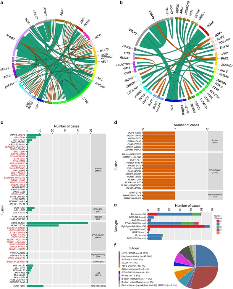
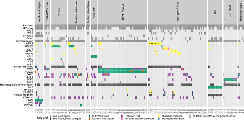
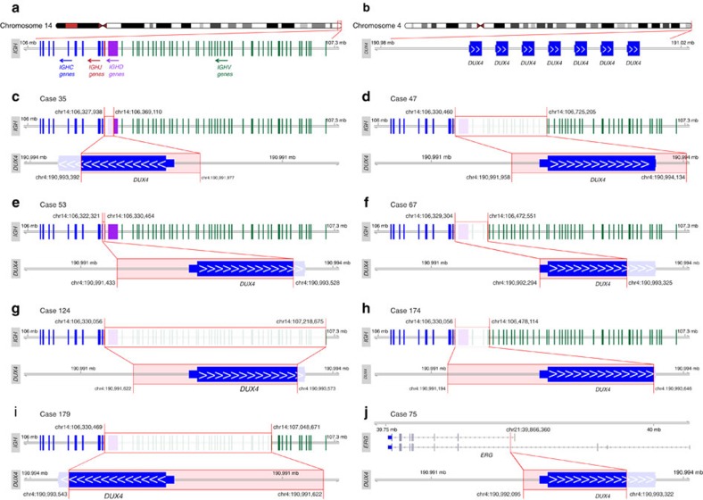
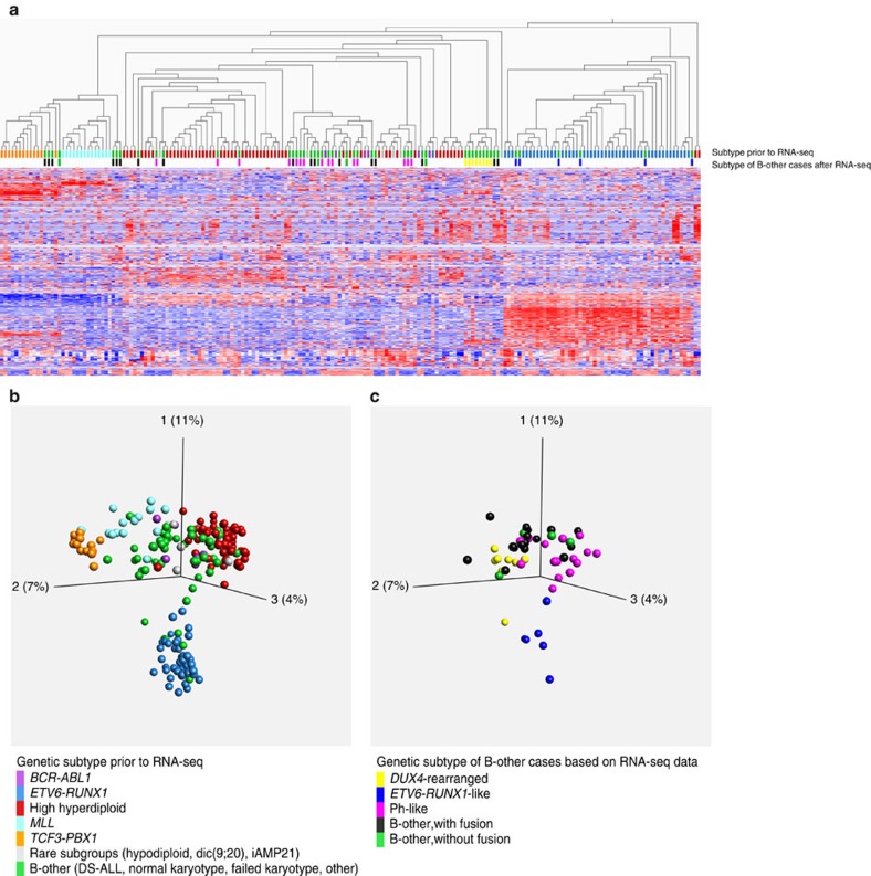
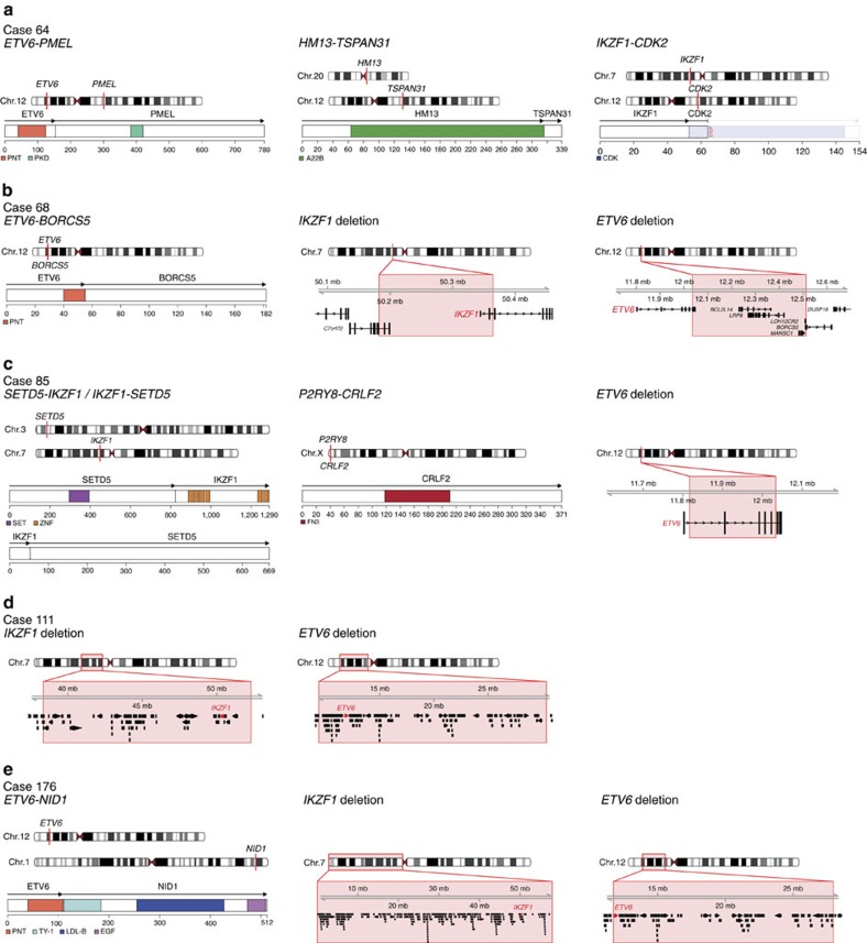
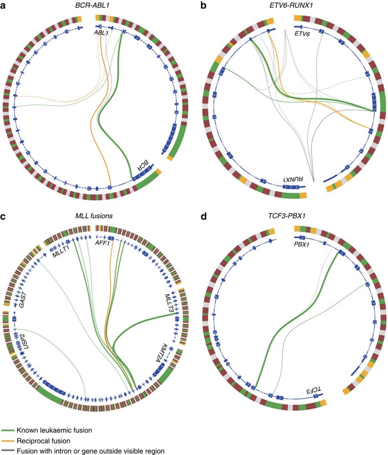
Fusion genes are potent driver mutations in cancer. In this study, we delineate the fusion gene landscape in a consecutive series of 195 paediatric B-cell precursor acute lymphoblastic leukaemia (BCP ALL). Using RNA sequencing, we find in-frame fusion genes in 127 (65%) cases, including 27 novel fusions. We describe a subtype characterized by recurrent IGH-DUX4 or ERG-DUX4 fusions, representing 4% of cases, leading to overexpression of DUX4 and frequently co-occurring with intragenic ERG deletions. Furthermore, we identify a subtype characterized by an ETV6-RUNX1-like gene-expression profile and coexisting ETV6 and IKZF1 alterations. Thus, this study provides a detailed overview of fusion genes in paediatric BCP ALL and adds new pathogenetic insights, which may improve risk stratification and provide therapeutic options for this disease.


Figures

Figure 1
Figure 1. Overview of the gene fusions present in 195 paediatric BCP ALL cases in the discovery cohort.
(a) In-frame gene fusions (green) and out-of-frame gene fusions (orange) are illustrated using Circos. Each ribbon has one end attached to the circle, indicating the 5′-partner gene of the fusion. The width of the ribbon is proportional to the number of detected fusions. Genes are arranged according to their genomic position (from chromosome 1–22 followed by X and Y) and chromosomes are marked in different colours. The gene symbol is denoted for genes involved in more than two unique fusions or in recurrent fusions. (b) In-frame gene fusions and out-of-frame gene fusions present in 50 B-other cases. The gene symbol for genes involved in more than two unique fusions or in recurrent fusions is indicated in bold. (c) The frequency of in-frame gene fusions by genetic subtype (indicated in the right column with the number of affected cases in parenthesis). Novel gene fusions are indicated in red (n=27, reciprocal gene-fusion pairs counted as a single fusion) and previously described fusions are indicated in black (n=22). (d) The frequency of out-of-frame gene fusions by genetic subtype (indicated in the right column with the number of affected cases in parenthesis). (e) Total number of gene fusions per case by genetic subtype (including both in-frame and out-of-frame fusions; reciprocal gene-fusion pairs counted as a single fusion). (f) Distribution of 195 BCP ALL cases within genetic subtypes defined by gene-expression profile and gene fusions detected by RNA-seq.
Figure 2
Figure 2. Genetic alterations present in 195 BCP ALL cases in the discovery cohort.
The cases are arranged according to genetic subtypes defined by gene-expression profile and gene fusions detected by RNA-seq, and were further characterized by SNP array, WGS, WES and MP-WGS. Genes recurrently altered in BCP ALL are arranged according to functional categories (kinase signalling, haematopoietic differentiation, histone modifiers and others). Events comprise induction failure and relapse.
Figure 3
Figure 3. DUX4 rearrangements in eight BCP ALL cases in the discovery cohort.
(a) Arrangement of immunoglobulin genes in the IGH locus. (b) Structure of the subtelomeric D4Z4 repeat region on 4q in the hg19 reference genome. This reference representation has seven repeats, each containing a DUX4 gene. Healthy individuals have 11–100 repeats. (c–i) Structure of the IGH-DUX4 rearrangements in (c) case 35, (d) case 47, (e) case 53, (f) case 67, (g) case 124, (h) case 174 and (i) case 179. (j) Structure of the ERG-DUX4 rearrangement in case 75. All genomic coordinates are based on the human reference genome hg19. Because it is impossible to determine which DUX4 repeat is involved in the rearrangement, the coordinates from the first DUX4 repeat are represented in the figures.
Figure 4
Figure 4. Hierarchical clustering and principal component analyses of RNA-seq gene-expression data.
A variance threshold was set at standard deviation 0.285, retaining 638 variables. The colour-coding of BCP ALL subtypes, used throughout the figure, is indicated in the bottom. (a) Unsupervised hierarchical clustering analysis of 195 BCP ALL cases. Coloured boxes below the dendogram indicate the subtype of each sample. The genetic subtype of B-other cases, based on the gene-expression and gene-fusion data, is indicated on the lower line. (b) Principal component analysis (PCA) of gene-expression data from all 195 BCP ALL cases. (c) PCA based on the data displayed in b, but only showing the 50 B-other cases colour-coded according to the genetic subtype based on the gene-expression and gene-fusion data. DS-ALL, Down's syndrome ALL; iAMP21, intrachromosomal amplification of chromosome 21.
Figure 5
Figure 5. Overview of aberrations in BCP ALL cases with ETV6-RUNX1-like gene-expression pattern.
The fusion genes are illustrated with a schematic overview of the chromosomal position of the genes involved in the fusion (top) and the retained protein domains together (bottom). Genomic deletions affecting ETV6 and IKZF1 are depicted with a red box indicating the deletion both at the chromosomal level and at the gene level, with ETV6 and IKZF1 in red. Illustrated protein domains: PNT, pointed domain; PKD, polycystic kidney disease domain; A22B, Peptidase A22B domain; CDK, protein kinase domain; SET, SET domain; ZNF, zinc-finger domain; FN3, fibronectin type-III domain; TY-1, thyroglobulin type-1 domain; LDL-B, LDL-receptor class B repeats; and EGF, EGF-like domain. (a) Gene fusions present in case 64. No SNP array data were available for this case. IKZF1-CDK2 is an out-of-frame fusion, with no functional domains from CDK2 being included in the fusion protein. (b) Gene fusions and deletions present in case 68. The breakpoints of the ETV6 deletion are within ETV6 and BORCS5; likely representing the event that created the ETV6-BORCS5 fusion gene. The breakpoints of the IKZF1 deletion occur within the C7orf72 and IKZF1 genes, but RNA-seq data did not indicate the presence of a fusion transcript of these genes. (c) Gene fusions and deletions present in case 85. The P2RY8-CRLF2 fusion does not contain any coding features from P2RY8 but leads to overexpression of the entire coding region of CRLF2. (d) Deletions present in case 111. No gene fusions were detected in this case. (e) Gene fusions and deletions present in case 176.
Figure 6
Figure 6. Splice patterns over fusion breakpoints.
Illustration of all detected fusion breakpoints in BCP ALL cases with (a) BCR-ABL1, (b) ETV6-RUNX1, (c) MLL fusions, and (d) TCF3-PBX1. Genes are arranged clockwise by genomic position. The outer circle represents the genomic region encompassing the indicated genes. Yellow indicates untranslated regions, green indicates coding exons, and red and grey indicate intronic regions (the latter are not to scale). The inner circle represents one or two overlaid reference transcripts of the indicated gene. Coding exons are indicated by a thick line with white arrows indicating the direction of the gene, introns are indicated by a thin or dashed line and untranslated regions are indicated by a medium thick line. Connecting lines between transcripts illustrate fusion breakpoints detected by at least three (for BCR-ABL1, ETV6-RUNX1 and MLL fusions) or ten reads (for TCF3-PBX1). Fusion breakpoints in individual BCP ALL cases are depicted in Supplementary Figs 15–18.

References

    1. Pui C.-H. & Evans W. E. Treatment of acute lymphoblastic leukemia. N. Engl. J. Med. 354, 166–178 (2006). - PubMed
    1. Martinelli G. et al.. IKZF1 (Ikaros) deletions in BCR-ABL1–positive acute lymphoblastic leukemia are associated with short disease-free survival and high rate of cumulative incidence of relapse: A GIMEMA AL WP report. J. Clin. Oncol. 27, 5202–5207 (2009). - PubMed
    1. Olsson L. et al.. Deletions of IKZF1 and SPRED1 are associated with poor prognosis in a population-based series of pediatric B-cell precursor acute lymphoblastic leukemia diagnosed between 1992 and 2011. Leukemia 28, 302–310 (2014). - PubMed
    1. Den Boer M. L. et al.. A subtype of childhood acute lymphoblastic leukaemia with poor treatment outcome: a genome-wide classification study. Lancet Oncol. 10, 125–134 (2009). - PMC - PubMed
    1. Mullighan C. G. et al.. Deletion of IKZF1 and prognosis in acute lymphoblastic leukemia. N. Engl. J. Med. 360, 470–480 (2009). - PMC - PubMed

Publication types

MeSH terms