Review
. 2016 Dec:41:10-18.
doi: 10.1016/j.sbi.2016.05.009. Epub 2016 Jun 5.

Structure and mechanism of assembly line polyketide synthases

Affiliations
Review

Structure and mechanism of assembly line polyketide synthases

Thomas Robbins et al. Curr Opin Struct Biol. 2016 Dec.

Abstract

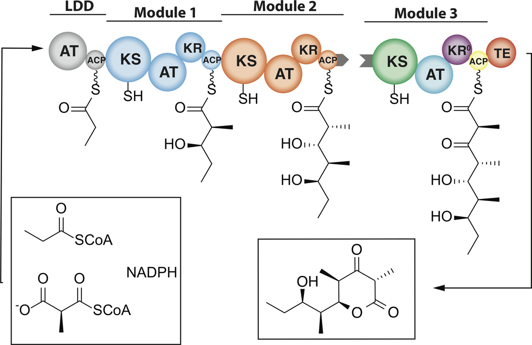
Assembly line polyketide synthases (PKSs) are remarkable biosynthetic machines with considerable potential for structure-based engineering. Several types of protein-protein interactions, both within and between PKS modules, play important roles in the catalytic cycle of a multimodular PKS. Additionally, vectorial biosynthesis is enabled by the energetic coupling of polyketide chain elongation to the channeling of intermediates between successive modules. A combination of high-resolution analysis of smaller PKS components and lower resolution characterization of intact modules and bimodules has yielded insights into the structure and organization of a prototypical assembly line PKS. This review discusses our understanding of key structure-function relationships in this family of megasynthases, along with a recap of key unanswered questions in the field.


Figures

Figure 1
Figure 1. A truncated three-module derivative of the 6-deoxyerythronolide B synthase (DEBS)
The first two PKS modules are shown in a distinct color, whereas the domains of Module 3 are colored for ease of comparison with the cryo-EM derived model of PikAIII (Figure 2A) and SAXS-derived model of DEBS Module 3 (Figure 2B). LDD = loading didomain; KS = ketosynthase; AT = acyltransferase; ACP = acyl carrier protein; KR = ketoreductase; KR0 = redox-inactive, epimerase-active ketoreductase; TE = thioesterase. Although the above assembly line PKS operates as a homodimer, for ease of visualizing the biosynthetic pathway, only one set of active sites are shown. Covalently linked domains are shown touching each other. The grey tabs refer to peptide docking domains that facilitate chain translocation between modules located in distinct polypeptide chains, such as Modules 2 and 3.
Figure 2
Figure 2. A.) Cryo-EM structure of PikAIII and B.) SAXS-derived model of DEBS Module 3
Equivalent domains are shown in the same color for ease of comparison between the two models and also with Figure 1.
Figure 3
Figure 3. The catalytic cycle of DEBS Module 2 (as depicted in Figure 1)
Step I: translocation of the diketide from the ACP domain of Module 1 onto the active site cysteine of the KS domain of Module 2. Step II: AT-catalyzed attachment of a methylmalonyl extender unit onto the Ppant arm of the ACP. Step III: Elongation of the KS-bound diketide chain by the ACP-bound methylmalonyl extender unit, followed by reduction of the ACP-bound (as depicted in Figure 1). Step I: translocation of the diketide from the ACP domain of Module 1 onto the active site cysteine of the KS domain of Module 2. Step II: AT-catalyzed attachmene triketide is translocated to Module 3.
Figure 4
Figure 4. KS-ACP docking models
Docking models are shown for Module 5 of DEBS. Panels A and C feature ACP4 (orange)-KS5 (green) interaction during chain translocation, whereas Panels B and D show ACP5 (yellow)-KS5 (green) interaction during chain elongation. Helix numbers are included for clarity. The models in panels A and B are based on the X-ray structure of the KS-AT fragment of DEBS Module 5 (PDB code: 2HG4), whereas those in panels B and D were derived by fitting individual domains into the cryo-EM maps (EMD-5651 and EMD-5653) of PikAIII.(Footnote)1 (Footnote)1 Homology models of ACP4 and ACP5 were generated using SWISS-MODEL[45] and the solution NMR structure of the ACP domain of DEBS Module 2 (PDB code: 2JU1). All docking models were generated using ClusPro server[46]. A Ppant group was added to both ACP domains in silico. A distance restraint of 10–20 A between active site residue of KS (C199) and the Ppant attachment site of ACP (S34) was introduced into each docking simulation. Additional experimental constraints used in this calculation are based on extensive mutagenesis data[38,39]. Top 20 models were superimposed and manually analyzed. Models that have lowest energy were selected as representatives. Rigid body fitting was performed using Chimera[47]. Real-space refinement with secondary structure restraints and group B-factor refinement was performed using PHENIX[48]. Figures were generated using PyMOL software (The PyMOL Molecular Graphics System, Version 1.7 Schrödinger, LLC).

References

    1. Walsh CT. Polyketide and Nonribosomal Peptide Antibiotics: Modularity and Versatility. Science. 2004;303:1805–1810. - PubMed
    1. Hopwood DA. Streptomyces in Nature and Medicine. 2007:1–261.
    1. Khosla C, Tang Y, Chen AY, Schnarr NA, Cane DE. Structure and Mechanism of the 6-Deoxyerythronolide B Synthase. Annu. Rev. Biochem. 2007;76:195–221. - PubMed
    1. Caffrey P, Bevitt DJ, Staunton J, Leadlay PF. Identification of DEBS 1, DEBS 2 and DEBS 3, the multienzyme polypeptides of the erythromycin-producing polyketide synthase from Saccharopolyspora erythraea. FEBS Journal. 1992;304:225–228. - PubMed
    1. Gokhale RS, Lau J, Cane DE, Khosla C. Functional Orientation of the Acyltransferase Domain in a Module of the Erythromycin Polyketide Synthase. Biochemistry. 1998;37:2524–2528. - PubMed

Publication types

MeSH terms