. 2016 Jun 7:16:170.
doi: 10.1186/s12906-016-1154-5.

High molecular weight of polysaccharides from Hericium erinaceus against amyloid beta-induced neurotoxicity

Affiliations

High molecular weight of polysaccharides from Hericium erinaceus against amyloid beta-induced neurotoxicity

Jai-Hong Cheng et al. BMC Complement Altern Med. .

Abstract

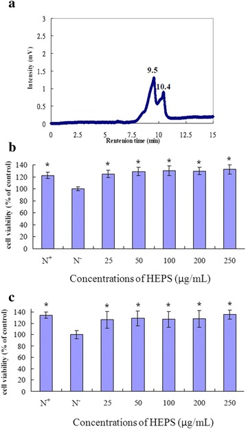
Background: Hericium erinaceus (HE) is a well-known mushroom in traditional Chinese food and medicine. HE extracts from the fruiting body and mycelia not only exhibit immunomodulatory, antimutagenic and antitumor activity but also have neuroprotective properties. Here, we purified HE polysaccharides (HEPS), composed of two high molecular weight polysaccharides (1.7 × 10(5) Da and 1.1 × 10(5) Da), and evaluated their protective effects on amyloid beta (Aβ)-induced neurotoxicity in rat pheochromocytoma PC12 cells.

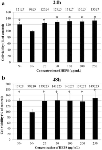
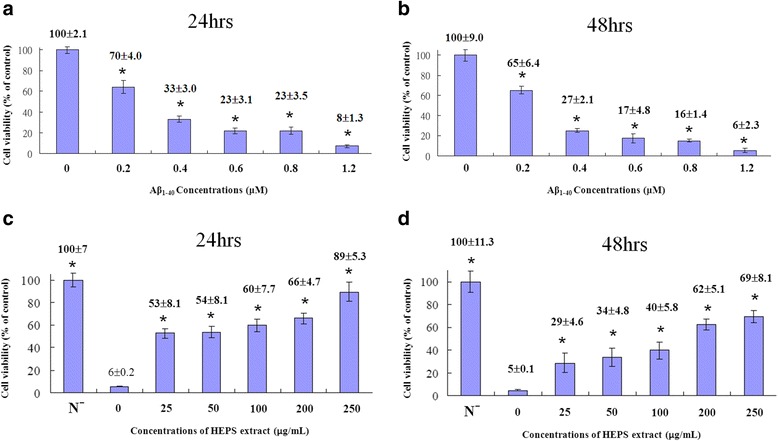
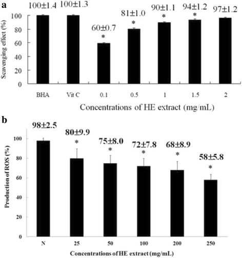
Methods: HEPS were prepared and purified using a 95 % ethanol extraction method. The components of HEPS were analyzed and the molecular weights of the polysaccharides were determined using high-pressure liquid chromatography (HPLC). The neuroprotective effects of the polysaccharides were evaluated through a 2,2-diphenyl-1-picrylhydrazyl (DPPH) radical scavenging assay and an MTT assay and by quantifying reactive oxygen species (ROS) and mitochondrial membrane potentials (MMP) of Aβ-induced neurotoxicity in cells.

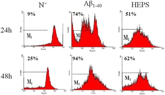
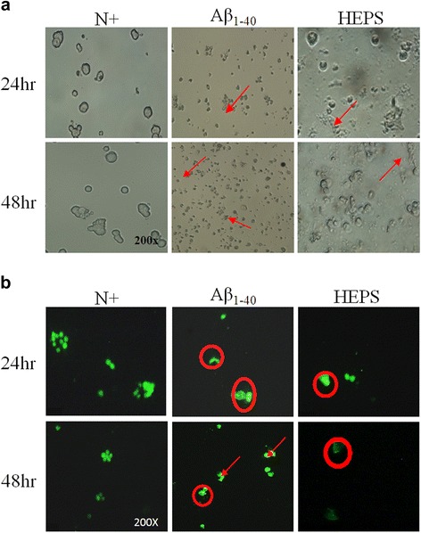
Result: Our results showed that 250 μg/ml HEPS was harmless and promoted cell viability with 1.2 μM Aβ treatment. We observed that the free radical scavenging rate exceeded 90 % when the concentration of HEPS was higher than 1 mg/mL in cells. The HEPS decreased the production of ROS from 80 to 58 % in a dose-dependent manner. Cell pretreatment with 250 μg/mL HEPS significantly reduced Aβ-induced high MMPs from 74 to 51 % and 94 to 62 % at 24 and 48 h, respectively. Finally, 250 μg/mL of HEPS prevented Aβ-induced cell shrinkage and nuclear degradation of PC12 cells.

Conclusion: Our results demonstrate that HEPS exhibit antioxidant and neuroprotective effects on Aβ-induced neurotoxicity in neurons.

Keywords: Amyloid beta; Hericium erinaceus; Neuroprotection; PC12 cell; Polysaccharides.


Figures

Fig. 1
Fig. 1
Measuring the molecular weight and toxicity of HEPS. a The molecular weight of HEPS was measured by HPLC. b 24 and (c) 48 h PC12 cell incubation with different concentrations of HEPS (25, 50, 100, 200 and 250 μg/mL, respectively). Cell viability was measured by an MTT assay with HEPS. RPMI-1640 complete medium containing 10 % horse serum and 5 % fetal bovine serum is indicated as N+. Serum-free RPMI-1640 medium is indicated as N. All values are mean ± SD and used a one-way analysis of variance (ANOVA, n = 6). All columns were significantly different compared with N and are indicated as * (p < 0.05)
Fig. 2
Fig. 2
The cell viability of PC12 cells with HEPS. a, (b) PC12 cells were treated with different concentrations (0, 25, 50, 100, 200 and 250 μg/mL) of HEPS and incubated for 24 and 48 h. Cell viability measurements were used to evaluate cell death using the MTT assay. Serum-free RPMI-1640 medium is indicated as N. All values are mean ± SD and used a one-way analysis of variance (ANOVA, n = 6). All columns were significantly different (p < 0.05)
Fig. 3
Fig. 3
The protective effects of HEPS on Aβ-induced toxicity in PC12 cells. a, (b) PC12 cells were treated with different concentrations of Aβ1–40 (0, 0.2, 0.4, 0.6, 0.8 and 1.2 μM) for 24 and 48 h and the toxicity was analyzed. c, (d) PC12 cells were treated with different concentrations (0, 25, 50, 100, 200 and 250 μg/mL) of HEPS followed by addition of 1.2 μM Aβ1–40 and incubation for 24 and 48 h. Cell viability measurements were used to evaluate cell death using the MTT assay. Serum-free RPMI-1640 medium is indicated as N. All values are mean ± SD and used a one-way analysis of variance (ANOVA, n = 6). All columns were significantly different (p < 0.05)
Fig. 4
Fig. 4
The scavenging activity of HEPS and production of ROS on Aβ-induced neurotoxicity in PC12 cells upon pretreatment of HEPS. a Different concentrations (0.1, 0.5, 1, 1.5 and 2 mg/mL) of HEPS were added into PC12 cells the free radical scavenging activity was measured using the DPPH assay. BHA and Vit C (0.1 mg/mL) were used as positive controls. b The effect of HEPS (25, 50, 100, 200 and 250 μg/mL) on the production of ROS was followed by 1.2 μM Aβ-induced neurotoxicity in PC12 cells. The production of ROS was measured using an ROS assay kit. N indicates samples without HEPS. All values are mean ± SD and used a one-way analysis of variance (ANOVA, n = 3 in panel A and n = 6 in panel B). All columns were significantly different (p < 0.05)
Fig. 5
Fig. 5
Measurement of MMP in PC12 cells. Cells were incubated with 1.2 μM Aβ1–40 for 24 and 48 h and MMP changes were measured using FACScan flow cytometry. MMPs were then reduced by pretreating samples with 250 μg/mL HEPS followed by 1.2 μM Aβ1–40. RPMI-1640 complete medium containing 10 % horse serum and 5 % fetal bovine serum is indicated as N+. Cells were incubated with 1.2 μM Aβ1–40 for 24 and 48 h, and MMP changes were measured using FACScan flow cytometry. MMPs were reduced by pretreating samples with 250 μg/mL HEPS followed by 1.2 μM Aβ1–40. RPMI-1640 complete medium containing 10 % horse serum and 5 % fetal bovine serum are indicated as N+
Fig. 6
Fig. 6
Morphological changes in PC12 cells were induced by Aβ, and cells were protected by pretreatment with HEPS. a Morphological changes in PC12 cells were observed by microscope after incubation with 1.2 μM Aβ1–40 for 24 and 48 h. b PC12 cells were protected by HEPS (250 μg/mL) followed by incubation with 1.2 μM Aβ1–40 for 24 and 48 h. Intracellular fluorescence was observed by fluorescence microscopy. RPMI-1640 complete medium containing 10 % horse serum and 5 % fetal bovine serum are indicated as N+. Circles and arrows indicate nuclear fragments and cell rupture, respectively

References

    1. Wang JC, Hu SH, Su CH, Lee TM. Antitumor and immunoenhancing activities of polysaccharide from culture broth of Hericium spp. Kaohsiung J Med Sci. 2001;17(9):461–7. - PubMed
    1. Sheu S-C, Lyu Y, Lee M-S, Cheng J-H. Immunomodulatory effects of polysaccharides isolated from Hericium erinaceus on dendritic cells. Process Biochem. 2013;48(9):1402–8. doi: 10.1016/j.procbio.2013.06.012. - DOI
    1. Yang BK, Park JB, Song CH. Hypolipidemic effect of an Exo-biopolymer produced from a submerged mycelial culture of Hericium erinaceus. Biosci Biotechnol Biochem. 2003;67(6):1292–8. doi: 10.1271/bbb.67.1292. - DOI - PubMed
    1. Wong KH, Sabaratnam V, Abdullah N, Kuppusamy UR, Naidu M. Effects of cultivation techniques and processing on antimicrobial and antioxidant activities of hericium erinaceus (Bull.:Fr.) Pers. Extracts. Food Technol Biotechnol. 2009;47(1):47–55.
    1. Lai PL, Naidu M, Sabaratnam V, Wong KH, David RP, Kuppusamy UR, et al. Neurotrophic properties of the Lion's mane medicinal mushroom, Hericium erinaceus (Higher Basidiomycetes) from Malaysia. Int J Med Mushrooms. 2013;15(6):539–54. doi: 10.1615/IntJMedMushr.v15.i6.30. - DOI - PubMed

MeSH terms

LinkOut - more resources