. 2016 Aug;160(2):473-83.
doi: 10.1016/j.surg.2016.04.014. Epub 2016 Jun 3.

Deficiency of cold-inducible ribonucleic acid-binding protein reduces renal injury after ischemia-reperfusion

Affiliations

Deficiency of cold-inducible ribonucleic acid-binding protein reduces renal injury after ischemia-reperfusion

Cindy Cen et al. Surgery. 2016 Aug.

Abstract

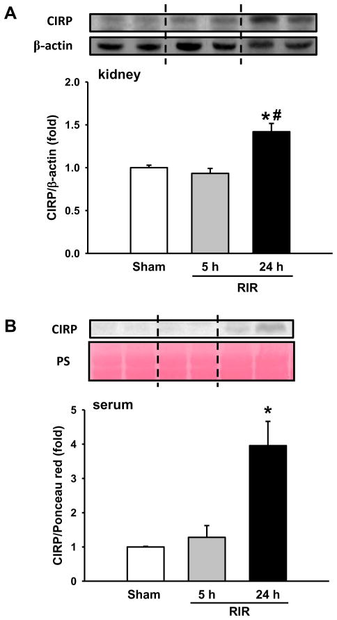
Background: Renal ischemia-reperfusion injury, commonly caused by major operation and shock, leads to acute kidney injury and is associated with high morbidity and mortality. Cold-inducible ribonucleic acid-binding protein, a cold shock protein, has recently been identified as a damage-associated molecular pattern. We hypothesized that cold-inducible ribonucleic acid-binding protein exacerbates severity of injury in renal ischemia-reperfusion.

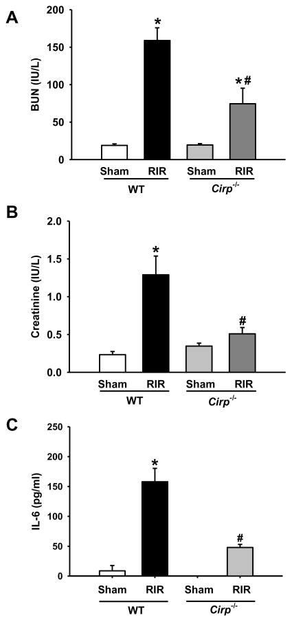
Methods: Renal ischemia was induced in an 8-week-old male C57BL/6 wild-type mice and Cirp(-/-) mice via bilateral clamping of renal pedicles for 30 minutes, followed by reperfusion for 5 or 24 hours and harvest of blood and renal tissue for analysis. Anti-cold-inducible ribonucleic acid-binding protein antibody or non-immunized immunoglobulin G (IgG) was injected intravenously (10 mg/kg body weight) at time of reperfusion.

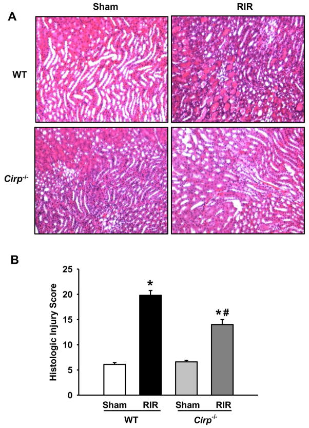
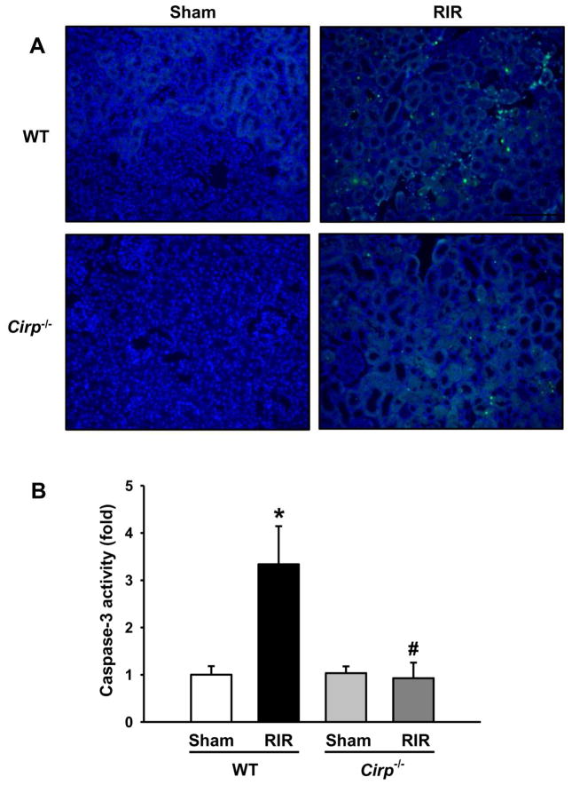
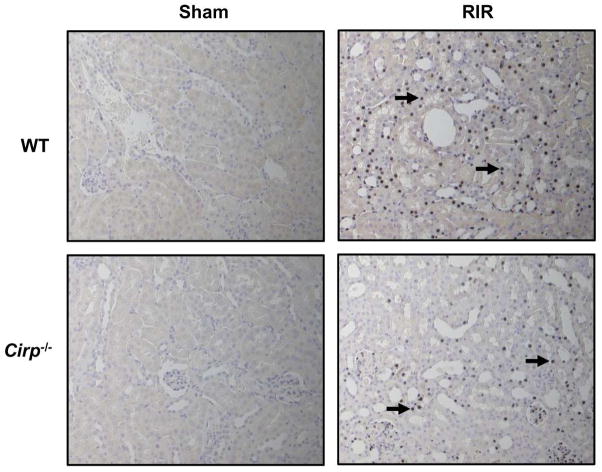
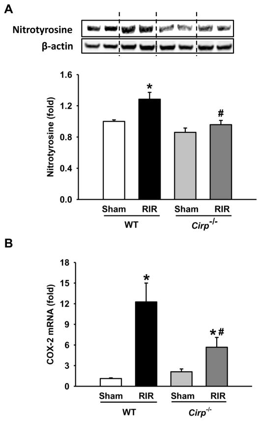
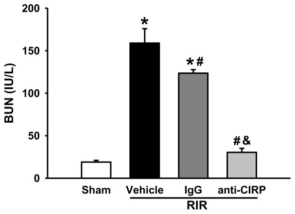
Results: After renal ischemia-reperfusion, Cirp(-/-) mice demonstrated a reduction of blood urea nitrogen and creatinine of 53% and 60%, respectively, compared with wild-type mice. Serum IL-6 levels were reduced significantly: 70% in Cirp(-/-) mice compared with wild-type mice after renal ischemia-reperfusion. Levels of nitrotyrosine, an oxidatively modified protein marker, and cyclooxygenase-2, an inflammatory mediator, also were significantly decreased in the kidneys of the Cirp(-/-) mice compared with wild-type mice after renal ischemia-reperfusion. Renal caspase-3 activity was decreased in Cirp(-/-) mice compared with wild-type mice after renal ischemia-reperfusion, which corresponded to the reduction of apoptotic cells determined by terminal deoxynucleotidyl transferase dUTP nick-end labeling assay. Injection of neutralizing anti-cold-inducible ribonucleic acid-binding protein antibody into wild-type mice led to an 82% reduction in blood urea nitrogen compared with the vehicle after renal ischemia-reperfusion.

Conclusion: Deficiency of cold-inducible ribonucleic acid-binding protein results in less renal injury after renal ischemia-reperfusion by attenuating inflammation and oxidative stress. Furthermore, blockade of cold-inducible ribonucleic acid-binding protein shows a protective effect, indicating cold-inducible ribonucleic acid-binding protein as a target in the treatment of renal ischemia-reperfusion.


Figures

Figure 1
Figure 1. CIRP expression after RIR
(A) Renal tissue was harvested at 5 and 24 h after 30 min-ischemia. The tissue was lysed and subjected to western blot against CIRP. Histogram shows mean densitometric analysis of protein bands after normalization with β-actin. (B) Western blot against CIRP in serum. n=5–9 mice/group. Data are expressed as mean ± SEM and compared by one-way ANOVA by SNK method. *p<0.05 vs WT sham; # p<0.05 vs WT vehicle.
Figure 2
Figure 2. Alterations in the levels of serum renal injury markers and cytokine after RIR
Serum was collected 24 h after 30 min-ischemia for measuring levels of (A) BUN and (B) creatinine. n=5–9 mice/group. Serum was also measured for levels of (C) IL-6 by ELISA. Sham, n=3–4 mice/group; RIR, n=5–6 mice/group. Data are expressed as mean ± SEM and compared by one-way ANOVA by SNK method. *p<0.05 vs WT sham; # p<0.05 vs WT vehicle.
Figure 3
Figure 3. Histological changes in WT and Cirp−/− kidneys after RIR
Renal tissue from sham WT and Cirp−/− mice and WT and Cirp−/− mice after RIR were harvested and subjected to histological analysis with hematoxylin-eosin staining. (A) Representative images were chosen from each group out of 10 randomly selected fields. Magnification 200×. (B) Histologic injury score measuring severity of tubular cell injury, tubular cell detachment, loss of brush border, tubular simplification, and cast formation on scale of 1–5 per category, as described in Materials and Methods. n=5–9 mice/group. Data are expressed as mean ± SEM and compared by one-way ANOVA by SNK method. *p<0.05 vs WT sham; # p<0.05 vs WT vehicle.
Figure 4
Figure 4. Apoptosis in the kidneys after RIR
Renal tissue from WT and Cirp−/− mice were harvested 24 h after 30 min-ischemia to evaluate for apoptosis. (A) Representative images of TUNEL staining (green fluorescence) and nuclear counterstaining (blue fluorescence). Magnification 200×. (B) Renal caspase-3 activity was determined spectrophotometrically. Sham, n=4 mice/group; RIR, n=6–7 mice/group. Data are expressed as means ± SEM and compared by one-way ANOVA by SNK method. *p<0.05 vs WT sham; # p<0.05 vs WT vehicle.
Figure 5
Figure 5. Neutrophil infiltration after renal I/R
Renal tissue from sham WT and Cirp−/− mice and WT and Cirp−/− mice after RIR were harvested and subjected to immunohistochemistry. Renal tissue was immunostained for Gr-1, a marker for neutrophil infiltration. Representative images shown for (A) WT sham, (B) WT RIR, (C) Cirp−/− sham, and (D) Cirp−/− RIR. Black arrows in (B) and (D) indicate representative staining for Gr-1. Magnification 200×.
Figure 6
Figure 6. Change in the levels of oxidatively-modified protein markers after RIR
Renal tissue from sham WT and Cirp−/− mice and WT and Cirp−/− mice after RIR were harvested. Tissue was lysed and subjected to western blot against (A) nitrotyrosine protein and β-actin. Histogram shows mean densitometric analysis of band intensities normalized to β-actin levels. Levels of (B) COX-2 mRNA were determined by real-time PCR. Sham, n=3–4 mice/group; RIR, n=4–7 mice/group. Data are expressed as means ± SEM and compared by one-way ANOVA by SNK method. *p<0.05 vs WT sham; # p<0.05 vs WT vehicle.
Figure 7
Figure 7. Alterations in serum organ injury marker BUN with anti-CIRP antibody after RIR
Mice were injected with anti-CIRP antibody or an IgG control after 30 min-ischemia time. Serum was collected 24 h later and assayed for levels of BUN. n=5–9 mice/group. Data are expressed as means ± SEM and compared by one-way ANOVA by SNK method. *p<0.05 vs sham; # p<0.05 vs vehicle; & p<0.05 vs IgG.

References

    1. Bonventre JV, Yang L. Cellular pathophysiology of ischemic acute kidney injury. J Clin Invest. 2011;121:4210–21. - PMC - PubMed
    1. Coca SG, Yusuf B, Shlipak MG, Garg AX, Parikh CR. Long-term risk of mortality and other adverse outcomes after acute kidney injury: a systematic review and meta-analysis. Am J Kidney Dis. 2009;53:961–73. - PMC - PubMed
    1. Munshi R, Hsu C, Himmelfarb J. Advances in understanding ischemic acute kidney injury. BMC Med. 2011;9:11. - PMC - PubMed
    1. Tian H, Sun T, Hao D, Wang T, Li Z, Han S, et al. The optimal timing of continuous renal replacement therapy for patients with sepsis-induced acute kidney injury. Int Urol Nephrol. 2014;46:2009–14. - PubMed
    1. Lopes JA, Jorge S, Resina C, Santos C, Pereira A, Neves J, et al. Acute kidney injury in patients with sepsis: a contemporary analysis. Int J Infect Dis. 2009;13:176–81. - PubMed

Publication types