. 2017 May 4;14(5):620-631.
doi: 10.1080/15476286.2016.1195940. Epub 2016 Jun 7.

Chromosome fusions triggered by noncoding RNA

Affiliations

Chromosome fusions triggered by noncoding RNA

John R Bracht et al. RNA Biol. .

Abstract

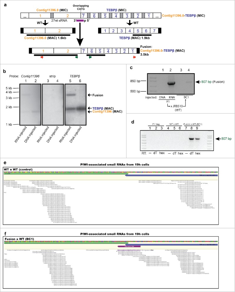
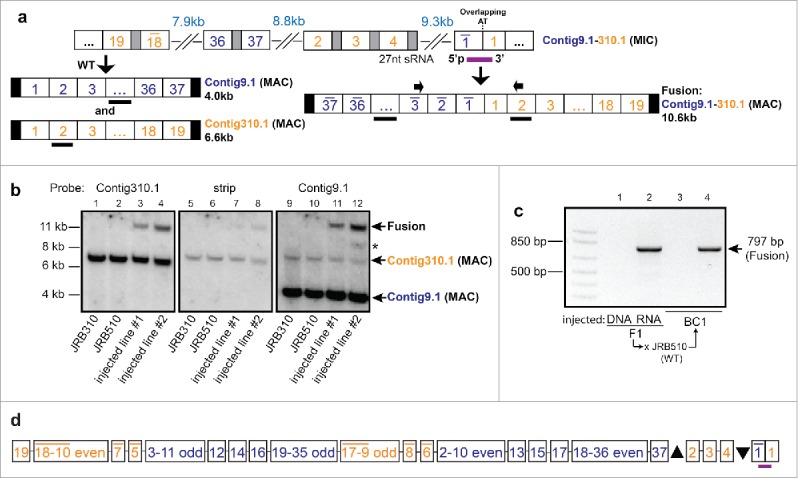
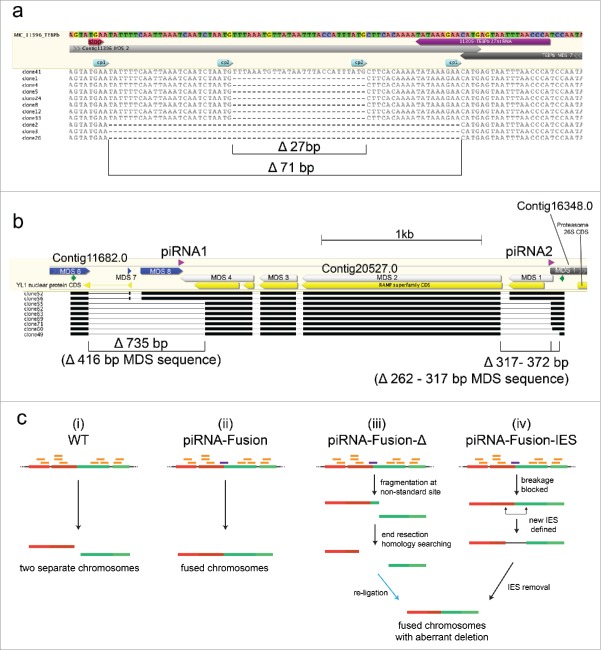
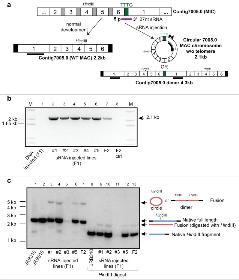
Chromosomal fusions are common in normal and cancer cells and can produce aberrant gene products that promote transformation. The mechanisms driving these fusions are poorly understood, but recurrent fusions are widespread. This suggests an underlying mechanism, and some authors have proposed a possible role for RNA in this process. The unicellular eukaryote Oxytricha trifallax displays an exorbitant capacity for natural genome editing, when it rewrites its germline genome to form a somatic epigenome. This developmental process provides a powerful model system to directly test the influence of small noncoding RNAs on chromosome fusion events during somatic differentiation. Here we show that small RNAs are capable of inducing chromosome fusions in 4 distinct cases (out of 4 tested), including one fusion of 3 chromosomes. We further show that these RNA-mediated chromosome fusions are heritable over multiple sexual generations and that transmission of the acquired fusion is associated with endogenous production of novel piRNA molecules that target the fused junction. We also demonstrate the capacity of a long noncoding RNA (lncRNA) to induce chromosome fusion of 2 distal germline loci. These results underscore the ability of short-lived, aberrant RNAs to act as drivers of chromosome fusion events that can be stably transmitted to future generations.

Keywords: Chromosome fusion; Oxytricha; long noncoding RNA; piRNAs.


Figures

Figure 1.
Figure 1.
Small RNA injection leads to heritable fusion of 2 somatic chromosomes encoding the non-scrambled genes, contig11396.0 and TEBPβ. (a) Schematic germline micronuclear (MIC, top) and somatic macronuclear (MAC, middle) maps are shown for each gene. The injected sRNA (purple bar), MDSs (numbered white boxes; TEBPβ MDS 1–7 are in the inverse orientation, indicated by a bar), IESs (gray boxes), somatic telomeres (black vertical rectangles), and a 4 bp overlap (CATG) between the 2 loci are not to scale. Locations of PCR primers are shown as small colored arrows; hybridization probes as thick black lines. (b) Southern analysis provides direct evidence for the presence of full-length somatic chromosome fusions in sRNA-injected but not DNA oligonucleotide-injected cells. “Strip” indicates an image of the stripped membrane before hybridizing to the TEBPβ probe. The full length WT TEBPβ chromosome is 1,858 bp, contig11396 is 1,635 bp and the fusion is predicted 3,493bp; each panel exposed to X-ray film for an equal time (24 hrs). (c) Transgenerational inheritance of the DNA fusion revealed by PCR analysis (with green primers) of a backcross (BC1) to WT strain JRB510. (d) RT-PCR with the same primers using oligo-dT (dT) or random hexamer (hex) primed cDNA from WT and fusion cells reveals conjugation-specific transcription (at 8–10 hrs) across the chromosomal fusion site, relative to asexually growing offspring of injected cells (Veg). (e) Mapping of piRNAs indicates an absence of piRNAs near the chromosomal fusion site in WT cells, but (f) the presence in BC1 cells of newly-produced piRNAs that bridge the normal chromosome ends but are distinct from the injected 27 nt sRNA (shown in purple). To normalize sequencing depth across libraries, the same number of raw, uncompressed reads (35 million) from each library were mapped onto the MIC contig containing TEBPβ and contig11396. Asterisks mark the 5′ ends of novel piRNAs and the injected sRNA (purple); contig11396, green; TEBPβ, blue.
Figure 2.
Figure 2.
Small RNA injection leads to heritable somatic fusion of 2 complex loci. (a) Fusion of 2 highly scrambled genes, contig9.1 and contig310.1, whose precursor MDS segments are intertwined in the germline on a 54 kb MIC contig (ctg7180000089708). Partial germline and somatic reference maps are shown, with segment numbers for contig9.1 in blue and contig310.1 in orange; other nomenclature as in Fig. 2 (full germline and somatic maps available: accession numbers given in Data Deposition section); PCR primers used to detect chromosomal fusion are indicated by small arrows above blue MDS 3 (inverted) and orange MDS 2; thick black bars denote Southern hybridization probes for contig9.1, spanning DNA segments 4–15, and for contig310.1 (MDS 2). (b) Southern analysis provides direct evidence for the presence of the full-length chromosome fusion induced by small RNA injection. Quantitative assessment of the phosphorimager signal in lane 4 (injected line 2 probed with contig310.1) suggests that the fusion chromosome is present at roughly half the levels of wild-type contig310.1. “Strip” is the signal remaining before hybridization to the Contig9.1 probe (exposure length and settings the same for each panel); asterisk indicates an aberrant band containing contig9.1 but not contig310.1. JRB310 and JRB510 are compatible WT mating strains of O. trifallax. (c) Transgenerational inheritance of the fusion chromosome revealed by PCR of a backcross to JRB510 cells (BC1, backcross generation 1). (d) Detailed map (not to scale) of the micronuclear locus containing intertwined precursor segments for these genes and 4 others. The upward pointing triangle represents 18 MDSs for 2 other genes (Contig15950 and Contig211.1), and the downward pointing triangle represents 20 MDSs for 3 other genes: Contig13252, Contig15950, and Contig7005.
Figure 3.
Figure 3.
Detection of aberrant deletions in some fusion products. (a) Deletions detected in the contig11396.0 - TEBPβ fusion. The injected piRNA is indicated in purple, and the clone sequence alignments are shown with dashes for deleted regions. “cp1” and “cp2” represent recombination between cryptic 3 bp pointers that resulted in 71 bp and 27 bp deletions, respectively. Only clone 41 is deletion-free. The stop codon (TGA) for the RAS homolog gene encoded on contig11396.0 is indicated in red. (b) Partial schematic representation of 3 neighboring germline loci and the locations of 2 co-injected small RNAs (purple). Cloning and sequencing of the PCR product between the 2 primers (small green arrows; gel shown in Fig. S1b) revealed fusion of all 3 somatic chromosomes in the progeny of injected cells. Partial germline structures of the 3 chromosomes (Contig11682.0, Contig20527.0, and Contig16348.0) are indicated in blue, light gray, and dark gray, respectively, with the complete gene and exon structures of these loci shown in Fig. S1c. Open reading frames are indicated in yellow (partial for the genes encoding YL1 Nuclear Protein and Proteasome 26S). Complex, combined deletions were observed in several sequenced clones. Black bars indicate sequence aligned regions, with thin black lines showing deleted regions. (c) A model for the formation of aberrant deletions during chromosome fusion. Two neighboring chromosomal loci are shown (indicated as red and green colored lines), each composed of 2 Macronuclear-Destined Segments (MDSs, indicated by slightly different shades of red or green). The injected piRNA is shown in purple and endogenous piRNAs in orange; thin black lines between MDSs indicate Internal Eliminated Sequences (IESs); thin dotted black lines mark flanking sequence. (i) Normal chromosome breakage in WT cells. (ii) piRNA-mediated chromosome fusion without deletions. (iii) piRNA mediated chromosome fusion with deletion. The re-ligation step is marked by a blue arrow. (iv) piRNA-mediated fusion containing a new IES-like deletion.
Figure 4.
Figure 4.
Small RNA injection leads to formation of a putative chromosome circle or dimer. (a) Schematic germline and somatic map of scrambled contig7005.0 and the injected sRNA (purple) that spans the end of the last MDS 6 and beginning of MDS 1, as well as a 4bp sequence that separates them; other nomenclature as in Fig. 2. The pair of inverse PCR primers used to detect the chromosome fusion are shown as small arrows; the thick black bar in MDS 1 denotes the Southern hybridization probe. (b) Inverse PCR confirms the formation and epigenetic inheritance of a fused chromosome in clonal lines derived from F1 and F2 cells. (c) Southern analysis of both undigested and HindIII digested total DNA provides direct evidence for the formation of the fusion in F1 progeny of sRNA-injected cells, as well as the offspring of a mixed mating between F1 lines #1-#5 (F2).
Figure 5.
Figure 5.
Microinjection of a long chimeric RNA leads to somatic formation of a hybrid TEBPβ/α chromosome. (a) Schematic map of injected RNA (1.375 kb): Gray and black horizontal bars denote Southern hybridization probes for TEBPβ and TEBPα, respectively; Numbered boxes are MDSs (not to scale); terminal black rectangles indicate telomeres; Locations of PCR primers to detect chimeric products are shown as colored arrows. (b) PCR confirms the formation of hybrid TEBPβ/α molecules in the progeny of sense (s) or antisense (as) RNA-injected cells but not uninjected cells (ctrl) (all primer and PCR sequences provided in Supplementary Information). (c) Southern analysis provides direct evidence for the presence of TEBPβ/α chimeric DNA molecules in the same cells used in (b). (d) Oligo-dT primed RT-PCR using either pair of primers detects chimeric RNA transcripts in the progeny of injected cells. Sequencing of the larger band confirmed that these RNA molecules do not contain the point substitution in the injected RNA. NT, no template control, RT+/− indicates the presence of reverse transcriptase enzyme; M, marker.

Similar articles

Cited by

References

    1. Sankar S, Lessnick SL. Promiscuous partnerships in Ewing's sarcoma. Cancer Genet 2011; 204:351-65; PMID:21872822; http://dx.doi.org/10.1016/j.cancergen.2011.07.008 - DOI - PMC - PubMed
    1. Mitelman F, Johansson B, Mertens F. The impact of translocations and gene fusions on cancer causation. Nat Rev Cancer 2007; 7:233-45; PMID:17361217; http://dx.doi.org/10.1038/nrc2091 - DOI - PubMed
    1. Heisterkamp N, Jenster G, Tenhoeve J, Zovich D, Pattengale PK, Groffen J. Acute-Leukemia in Bcr/Abl Transgenic Mice. Nature 1990; 344:251-3; PMID:2179728; http://dx.doi.org/10.1038/344251a0 - DOI - PubMed
    1. Adams JM, Harris AW, Pinkert CA, Corcoran LM, Alexander WS, Cory S, Palmiter RD, Brinster RL. The C-Myc Oncogene Driven by Immunoglobulin Enhancers Induces Lymphoid Malignancy in Transgenic Mice. Nature 1985; 318:533-8; PMID:3906410; http://dx.doi.org/10.1038/318533a0 - DOI - PubMed
    1. Daley GQ, Vanetten RA, Baltimore D. Induction of Chronic Myelogenous Leukemia in Mice by the P210bcr/Abl Gene of the Philadelphia-Chromosome. Science 1990; 247:824-30; PMID:2406902; http://dx.doi.org/10.1126/science.2406902 - DOI - PubMed

Publication types