Review
. 2016 Aug;37(8):625-640.
doi: 10.1016/j.tips.2016.05.006. Epub 2016 Jun 4.

Recent Structural Insights into Cytochrome P450 Function

Affiliations
Review

Recent Structural Insights into Cytochrome P450 Function

F Peter Guengerich et al. Trends Pharmacol Sci. 2016 Aug.

Abstract

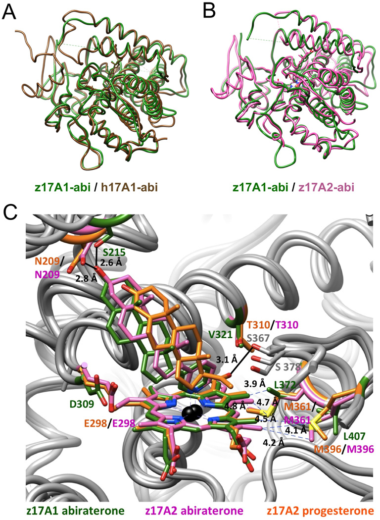
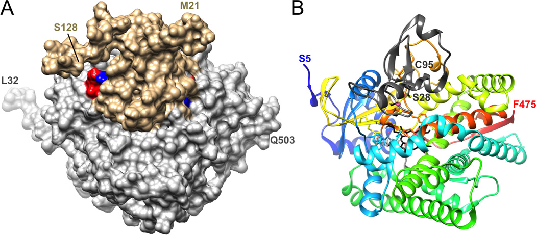
Cytochrome P450 (P450) enzymes are important in the metabolism of drugs, steroids, fat-soluble vitamins, carcinogens, pesticides, and many other types of chemicals. Their catalytic activities are important issues in areas such as drug-drug interactions and endocrine function. During the past 30 years, structures of P450s have been very helpful in understanding function, particularly the mammalian P450 structures available in the past 15 years. We review recent activity in this area, focusing on the past 2 years (2014-2015). Structural work with microbial P450s includes studies related to the biosynthesis of natural products and the use of parasitic and fungal P450 structures as targets for drug discovery. Studies on mammalian P450s include the utilization of information about 'drug-metabolizing' P450s to improve drug development and also to understand the molecular bases of endocrine dysfunction.

Keywords: antibiotic discovery; cytochrome P450; drug metabolism; natural product biosynthesis; steroid metabolism.


Figures

Figure 1
Figure 1
Tertiary structure of the P450 21A2 progesterone complex and active site configuration (PDB 4Y8W; www.rcsb.org) [28]. (A) Ribbon diagram of the overall fold of P450 21A2 with rainbow coloring from blue (N-terminus) to red (C-terminus). Individual secondary structural elements are labeled; carbon atoms of heme and progesterone are colored in gray; Fe3+ is shown as an orange sphere. Membrane P450s feature at least 12 α-helices that are designated with letters A to L and an N-terminal β-sheet domain with consecutive numbering of strands [27]. The protein core comprises helices C, D, I, K, and L and the β-sheets that make up part of the heme-binding site and the adjacent region where partner proteins dock. Substrates bind in a cleft above the heme, with several channels opening to the surface. The catalytic cysteine is located on the other side of heme, such that its thiolate moiety occupies the axial coordination site of iron opposite the bound oxygen. Helices B, C, F, and G that are situated more on the periphery of the protein and form the outer boundaries of the substrate cavity exhibit more structural variations and can be expected to display a more dynamic behavior as do the N-and C-terminal ends. (B) Close-up view of the active site with progesterone surrounded by Fourier 2Fo-Fc omit electron density drawn at the 1.2 σ threshold. Selected amino acids and α-helices wrapping around heme or forming the ceiling of the active site are labeled. All images were generated with the program UCSF Chimera [29].
Figure 2
Figure 2
Crystal structure and active site volume of bacterial P450revI (PDB 3WVS) [36]. Ribbon diagram of P450revI with rainbow coloring from blue (N-terminus) to red (C-terminus). The active site with a very large volume of ~2500 Å3 (the difference between the gray and magenta bodies, the latter occupied by heme that is part of the enzyme) is shown as a semi-transparent surface, and heme and reveromycin T are highlighted in ball-and-stick mode with carbon atoms colored in magenta and black, respectively.
Figure 3
Figure 3
Binding of two ligands in the crystal structures of the human P450:progesterone (PDB 4Y8W) [49] and bovine P450 21A2:17α-hydroxyprogesterone (PDB 3QZ1) [60] complexes. (A) Superimposition of the structures of the human (green) and bovine (pink) complexes with ligands bound at the canonical active site (progesterone - stick mode with black bonds, or 17α-hydroxyprogesterone – ball-and-stick mode with carbon atoms in gray) and a distal site (17α-hydroxyprogesterone) indicated. (B) Close-up view of the superimposed distal site regions in the human and bovine complex structures, with Fourier 2Fo-Fc sum electron density from the human structure (green meshwork, 0.7σ threshold) indicative of binding of progesterone of partial occupancy at a location that matches that of the distal site 17α-hydroxyprogesterone in the bovine structure. Selected helices and amino acids are labeled.
Figure 4
Figure 4
Crystal structures of P450 105AS1 ligand-free and with ligand bound (PDB 4OQR and 4OQS, respectively) [80]. Superimposition of the ligand-free structure of P450 105AS1 (beige ribbon and heme) and the structure of the complex with compactin (cyan ribbon and heme, and ligand depicted in ball-and-stick mode with carbon atoms colored in magenta). The comparison reveals the reorientation of the B-C loop region (left) and movements by the F and G helices (top).
Figure 5
Figure 5
Similar tertiary structure folds of human P450 17A1 and zebrafish P450 17A1/A2 and subtle differences in the active sites of zebrafish P450 17A1 and 17A2 [103]. (A) Overlay of the abiraterone (abi) complexes of human P450 17A1 (PDB 3RUK; brown) [84] and zebrafish 17A1 (PDB 4R1Z; green). (B) Overlay of the abiraterone complexes of zebrafish P450 17A1 (green) and P450 17A2 (PDB 4R20; pink). (C) Comparison between the active site configurations of the zebrafish P450 17A1:abiraterone (green), 17A2:abiraterone (pink) and 17A2:progesterone (PDB 4R21; orange) complexes reveals five differences in the amino acid compositions of 17A1 (from human or zebrafish) and 17A2 (from zebrafish) in the vicinity of the heme moiety and near the ceiling of the active site. Color labels of residues match the corresponding structures.
Figure 6
Figure 6
Three-dimensional structures of P450 complexes. (A) Surface representation of the complex between human P450 17A1 (gray) and cytochrome b5 (beige) based on cross-linking and mass spectrometric analysis [106]. The model was generated with the program HADDOCK [108], using the X-ray coordinates of human 17A1 (PDB 3RUK) [100] and NMR coordinates of human b5 (PDB 2I96). Two salt bridges identified by XLMS are highlighted in blue (lysine) and red (glutamate), and N- and C-terminal residues of both proteins are labeled. (B) Crystal structure of the complex between P450 11A1 and adrenodoxin (Adx, PDB 3N9Y) [107]. P450 11A1 is shown in cartoon mode with rainbow coloring from blue (N-terminus) to red (C-terminus), adrenodoxin is depicted as a dark gray ribbon (only a portion of Adx, residues Ser-28 to Cys-95, was visible in the crystal structure), carbon atoms of heme and cholesterol are colored in black and gray, respectively, and iron (P450 Fe3+ and Adx [2Fe-2S] cluster) and sulfur atoms are highlighted as pink and yellow spheres, respectively. The complexes depicted in the two panels are shown in similar orientations.

References

    1. Omura T, Sato R. A new cytochrome in liver microsomes. J. Biol. Chem. 1962;237:1375–1376. - PubMed
    1. Mueller GC, Miller JA. The metabolism of 4-dimethylaminoazobenzene by rat liver homogenates. J. Biol. Chem. 1948;176:535–544. - PubMed
    1. Ryan KJ. Conversion of androstenedione to estrone by placental microsomes. Biochim. Biophys. Acta. 1958;27:658–662. - PubMed
    1. Gillette JR, Brodie BB, La Du BN. The oxidation of drugs by liver microsomes: on the role of TPNH and oxygen. J. Pharmacol. Exp. Therapeut. 1957;119:532–540. - PubMed
    1. Rendic S, Guengerich FP. Survey of human oxidoreductases and cytochrome P450 enzymes involved in the metabolism of xenobiotic and natural chemicals. Chem. Res. Toxicol. 2015;28:38–42. - PMC - PubMed

Publication types

MeSH terms

Substances