. 2016 Jun 7;9(1):63.
doi: 10.1186/s13041-016-0243-1.

Microvascular endothelial cells-derived microvesicles imply in ischemic stroke by modulating astrocyte and blood brain barrier function and cerebral blood flow

Affiliations

Microvascular endothelial cells-derived microvesicles imply in ischemic stroke by modulating astrocyte and blood brain barrier function and cerebral blood flow

Qunwen Pan et al. Mol Brain. .

Abstract

Background: Endothelial cell (EC) released microvesicles (EMVs) can affect various target cells by transferring carried genetic information. Astrocytes are the main components of the blood brain barrier (BBB) structure in the brain and participate in regulating BBB integrity and blood flow. The interactions between ECs and astrocytes are essential for BBB integrity in homeostasis and pathological conditions. Here, we studied the effects of human brain microvascular ECs released EMVs on astrocyte functions. Additionally, we investigated the effects of EMVs treated astrocytes on regulating BBB function and cerebral ischemic damage.

Results: EMVs prepared from ECs cultured in normal condition (n-EMVs) or oxygen and glucose deprivation (OGD-EMVs) condition had diverse effects on astrocytes. The n-EMVs promoted, while the OGD-EMVs inhibited the proliferation of astrocytes via regulating PI3K/Akt pathway. Glial fibrillary acidic protein (GFAP) expression (marker of astrocyte activation) was up-regulated by n-EMVs, while down-regulated by OGD-EMVs. Meanwhile, n-EMVs inhibited but OGD-EMVs promoted the apoptosis of astrocytes accompanied by up/down-regulating the expression of Caspase-9 and Bcl-2. In the BBB model of ECs-astrocytes co-culture, the n-EMVs, conversely to OGD-EMVs, decreased the permeability of BBB accompanied with up-regulation of zonula occudens-1(ZO-1) and Claudin-5. In a transient cerebral ischemia mouse model, n-EMVs ameliorated, while OGD-EMVs aggravated, BBB disruption, local cerebral blood flow (CBF) reduction, infarct volume and neurological deficit score.

Conclusions: Our data suggest that EMVs diversely modulate astrocyte functions, BBB integrity and CBF, and could serve as a novel therapeutic target for ischemic stroke.

Keywords: Astrocytes; Blood brain barrier; Cerebral blood flow; Cerebral ischemia; Endothelial cells; Gene expression; Microvesicles.


Figures
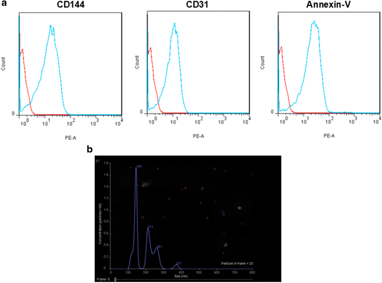
Fig. 1
Fig. 1
Characterization of EMVs by flow cytometric analysis and NTA. a Flow cytometric analysis showing the expression of MV specific marker (Annexin V) and endothelial cell specific markers (CD31 and CD144) in EMVs. b NTA analysis confirmed the size distribution of EMVs
Fig. 2
Fig. 2
The incorporation of EMVs with astrocytes after coculture (A) Representative images showing that EMVs merged with astrocytes. EMVs were labeled with PKH26 (red). Nucleuses were labeled with DAPI (blue). Scale bar, 50 μm
Fig. 3
Fig. 3
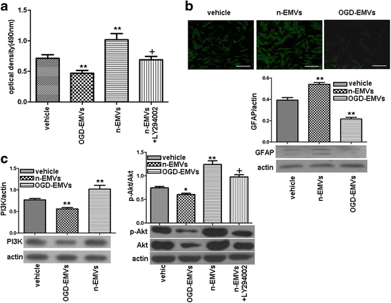
The effects of n-EMVs and OGD-EMVs on astrocyte proliferation and expression of GFAP and PI3K/Akt were opposite. a MTT assay of astrocyte proliferation. b Representative images and quantitative analysis of GFAP in each group. Scale bar, 50 μm. c Expression of PI3K and p-Akt/Akt. (** p <0.01,vs. vehicle; + p <0.05,vs. n-EMVs, n = 5)
Fig. 4
Fig. 4
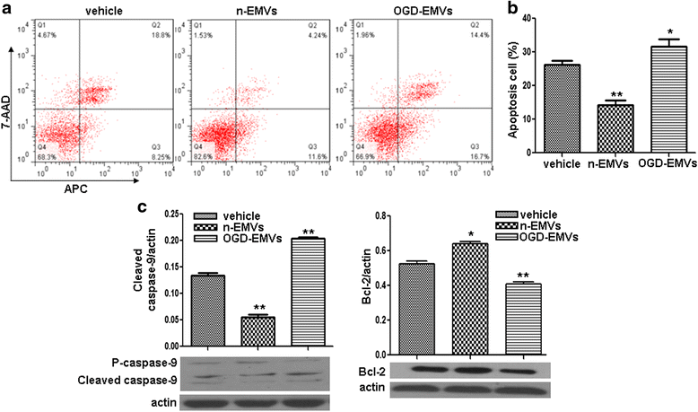
The effects of n-EMVs and OGD-EMVs on astrocyte apoptosis and expression of cleaved Caspase-9 and Bcl-2. a Representative flow cytometric analysis of astrocytes apoptosis. b Summarized data on the percentage of apoptotic astrocytes in each group. c Cleaved Caspase-9 and Bcl-2 expression in astrocytes. (* p <0.05,** p <0.01,vs. vehicle, n = 5)
Fig. 5
Fig. 5
Treatment with n-EMVs and OGD-EMVs reversely influenced the permeability of BBB and ZO-1/Claudin-5 expression. a n-EMVs reduced while OGD-EMVs increased the permeability of BBB. b, c Representative images of ZO-1 (b) and Claudin-5 (c) staining on HBMECs. Scale bar, 50 μm. d Western blot analyses of ZO-1 and Claudin-5. (* p <0.05, ** p <0.01, vs. vehicle, n = 5)
Fig. 6
Fig. 6
Representative images showing that injected EMVs merged with astrocytes in the peri-infarct area. EMVs labeled with PKH26 (red), and astrocytes labeled with GFAP (green). Nucleuses stained with DAPI (blue). Magnification, 400×
Fig. 7
Fig. 7
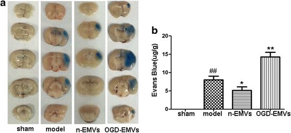
N-EMVs reduced while OGD-EMVs promoted evans blue extravasation in tMCAO mice. a The representative pictures of Evans blue extravasation in ischemic right brains of various groups. b The quantitative analysis of Evans blue leakage. (* p <0.05, ** p <0.01,vs. model group, n = 5)
Fig. 8
Fig. 8
Effects of EMVs on infract area, neurological deficits score and CBF. ab Representative brain TTC staining and quantitative analysis of infarct size in different groups. cd The representative images and analysis of CBF in different groups. Blue to red represent low to high perfusion. e Neurological deficits score in different groups. (* P <0.05, ** p <0.01 vs. model group, n = 5)

References

    1. Zhu H, Wang Z, Xing Y, Gao Y, Ma T, Lou L, et al. Baicalin reduces the permeability of the blood–brain barrier during hypoxia in vitro by increasing the expression of tight junction proteins in brain microvascular endothelial cells. J Ethnopharmacol. 2012;141(2):714–20. doi: 10.1016/j.jep.2011.08.063. - DOI - PubMed
    1. Shah K, Abbruscato T. The role of blood–brain barrier transporters in pathophysiology and pharmacotherapy of stroke. Curr Pharm Des. 2014;20(10):1510–22. doi: 10.2174/13816128113199990465. - DOI - PubMed
    1. Densmore JC, Signorino PR, Ou J, Hatoum OA, Rowe JJ, Shi Y, et al. Endothelium-derived microparticles induce endothelial dysfunction and acute lung injury. Shock. 2006;26(5):464–71. doi: 10.1097/01.shk.0000228791.10550.36. - DOI - PubMed
    1. Jung JE, Kim GS, Chen H, Maier CM, Narasimhan P, Song YS, et al. Reperfusion and neurovascular dysfunction in stroke: from basic mechanisms to potential strategies for neuroprotection. Mol Neurobiol. 2010;41(2–3):172–9. doi: 10.1007/s12035-010-8102-z. - DOI - PMC - PubMed
    1. Strazielle N, Ghersi-Egea JF. Physiology of blood–brain interfaces in relation to brain disposition of small compounds and macromolecules. Mol Pharm. 2013;10(5):1473–91. doi: 10.1021/mp300518e. - DOI - PubMed

MeSH terms